Download
1 / 26
260 likes | 514 Views
BREASTS. Most prominent superficial structures in the ant thoracic wall, especially in women Consists of: Glandular tissue Supporting fibrous tissue Fatty matrix Blood vessels Nerves Lymphatics. BREASTS. Mammary glands

E N D
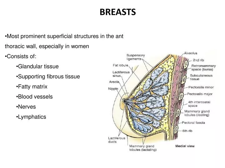
BREASTS • Most prominent superficial structures in the ant thoracic wall, especially in women • Consists of: • Glandular tissue • Supporting fibrous tissue • Fatty matrix • Blood vessels • Nerves • Lymphatics
BREASTS • Mammary glands • in subcutaneous tissue overlying pectoralis major and minor muscles • accessory to reproduction in women • Greatest prominence = nipple • Circular pigmented area of skin surrounding the nipple = areola
MALE BREASTS • In Men: • Mammary glands are rudimentary and function-less • few small ducts or epithelial cords • Fat same as subcutaneous tissue found elsewhere • Glandular system does not develop
FEMALE BREAST • Consists of glandular tissue (parenchyma) and connective tissue (stroma) • Amount of fat surrounding glandular tissue determines size of non-lactating breast • Breast on bed that extends transversely from lat border of sternum to midaxillary line and vertically from 2nd – 6th ribs • 2/3 of bed formed by pectoralis fascia (overlying pectoralis major) • 1/3 formed by fascia covering serratus ant
Retromammary space – between breast and pectoral fascia • loose connective tissue plane or potential space • contains small amount of fat • allows movement of breast on pectoral fascia
Glandular component (mammary gland) consists of lobes separated by fat and suspensory ligaments • Lobes subdivided by lobules (functional component of mammary gland) which contains alveoli • Alveoli lined by milk secreting cuboidal cells surrounded by myoepithelial cells • Lobules and lobes drained by a lactiferous duct – carries milk to nipple • Ducts has dilated portion deep to areola, lactiferous sinus – milk accumulates • Ducts are blocked by keratin plug in non-lactating women – mechanical barrier which prevents bacteria from entering the duct
Axillary process or tail of Spence - small part of mammary gland may extend along infro-lateral edge of pectoralis major toward axillaryfossa through the deep fascia • Lobes bound together by interlobular connective tissue • Suspensory ligaments (of Cooper) extend from interlobular connective tissue to attach mammary glands to dermis of overlying skin • Support lobes and lobules of glands
Areola – contains numerous sebaceous glands • areolar glands of Montgomery • enlarge during pregnancy • secrete an oily substance – protective lubricant for areola and nipple • Nipples – 4th ICS, lateral to midclavicular line • are fissured with the lactiferous ducts opening into them • composed of smooth muscle fibers which compresses lactiferous ducts during lactation and erect nipples in response to stimulation (baby begins to suckle)
Arterial supply • Medial mammary branches of perforating branches of internal thoracic art • Ant intercostal branches of internal thoracic art • (internal thoracic art branch of 1st part of subclavian artery) • Lateral thoracic (lat mammary art) and thoracoacromial arteries (pectoral branch) of axillary artery • Post intercostal arteries – branches of thoracic aorta of 2nd, 3rd and 4th ICS
Venous drainage • Mainly to axillary vein • Also some drainage to internal thoracic vein
Lymphatic drainage • NB because of role in metastasis of cancer cells • Nipple, areola, lobules of glands • Subareolar lymphatic plexus • Axillary LN parasternal LN or opposite breast >75% (especially lat quadrant) Most of remaining lymph (especially from medial quadrant) abd LN (subdiaphragmaticinfphrenic LN) Inf quadrant
Lymphatic drainage • Axillary LN • Pectoral • Subscapular • Apical • Humoral • Central • Initially to pectoral but may go directly to other axillary LNs • Can even drain • Interpectoral • Deltopectoral • Inf deep cervical • supraclavicular
Lymphatic drainage • Skin of the breast • Ipsilataxillary LN • Inf deep cervical LN • Infraclavicular LN • Parasternal LN of both sides
Lymphatic drainage • Axillary LN • Clavicular • (infra- and supraclavicular LN) • Subclavian lymphatic trunk • (also drains from the upper limb)
Lymphatic drainage • Parasternal LN • Bronchomediastinal lymphatic trunk • (also drains thoracic viscera)
Subclavian lymphatic trunk • Bronchomediastinal lymphatic trunk • Merge with jugular lymphatic trunk • (drains head and neck) • Form lymphatic duct on R • OR • Enters thoracic duct on L • OR • Open independently in R or L venous angle • ( junction of internal jugular and subclavian veins to form brachiocephalic veins Lymphatic drainage
Nerve supply • Ant and lat cutaneous branches of 4th-6th IC nerves • Sensory fibers to skin • Sympathetic fibers to • Blood vessels • Smooth muscle of overlying skin and nipple
EMBRYOLOGY OF THE MAMMARY GLAND • Embryo has 3 distinguishable layers of cells which give rise to various tissues • Ectoderm • Mesoderm • Endoderm • +/- 32 days broad band of ectodermal cells form on either side of trunk from upper limb to lower limb = mammary band • mammary streak • Mammary lines (4th-5th week) Mammary gland Connective tissue Ectodermal differentiation in ventro-lat aspect of body wall
mammary lines • Mammary crest (at 4 weeks) • Mammary Hillock • Mammary bud (at 6 weeks) Shorten Ectodermal cells divide and grow into mesenchymal cell layer Proliferate to form a ridge Ridge disappears Only remain pectoral region Ectodermal layer continue to grow in mesenchymal cell layer Ectodermal cells continue to grow in mesenchymal cell layer forming a globular structure Primordium of gland
Rapid growth of mesenchyme around mammary bud • Blood vessels and connective tissue develop from mesenchyme or mesoderm that surrounds the buds and organise developing buds into lobules • Invagination of mammary bud cells into mesenchyme • Becomes the primary bud (gives rise to gland cistern)
12th week – epidermis becomes depressed – shallow mammary pit from which nipples arise because of proliferation of surrounding connective tissue of the areola
Development of mesenchyme very sensitive for testosterone for short while (15 weeks) • Too much testosterone – increased proliferation of mesenchyme • Stop development of buds • Arrest in breast development • Baby born without breasts • at 5 months, surface of mammary bud spreads out and depression form • Deep layer of bud epithelium proliferates and produces secondary buds
Secondary buds lengthen and become solid cords of epithelial cells growing into mesenchymal tissue • Will form the lactiferous ducts • Canalisation – process of forming a lumen in solid core of epithelial cells • 20th-32nd week • will continue until canals are lined with only a couple of layers of epithelial cells • Thus up to 40th week buds develop from lactiferous ducts and lobular-alveolar system is formed
After birth: • Organ is rudimentary up to puberty • Puberty development further takes place in females • Growth of duct system and fat deposits in lobules • In pregnancy: • Further development takes place • 1st half – duct system develop • 2nd half – alveolar compartment develop further • Lactation: • Development optimal • Organ fully functional
ABNORMALITIES Disappearance of mammary crest or failure of development – absent nipples and breasts Failure of mammary bud to form – absent nipples and breasts