Download
1 / 1
10 likes | 110 Views
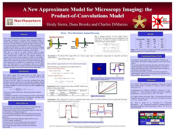
There has been increasing interest in recent years in techniques for microscopic examination of optically thick transparent objects. A need for 3D imaging models for phase microscopes that allow deconvolution techniques has emerged. In response, a Product-of-Convolutions (POC) model was developed to maintain accuracy for thick objects while being computationally tractable. This model compares the Born, Rytov, and Product models in a common framework. The availability of a tractable accurate computational model would aid instrument design and quantitative reconstruction. The POC model outperforms the Born and Rytov approximations, particularly for thick objects. Future work will include proving that the POC model corresponds to an approximation solution of the wave equation.

E N D
Expected Product Rytov Born Results Abstract There has been increasing interest in recent years in techniques for microscopic examination of optically thick transparent objects. A number of phase imaging modalities have been developed to address this need. If a stack of images is acquired through focusing, the image at a given focal plane is contaminated by out-of-focus information coming from other planes [1]. There is a need to develop 3D imaging models for phase microscopes that will allow deconvolution, or more generally inverse reconstruction, techniques to be developed. Thus there is a need for an image formation model for phase microscopy that is able to maintain accuracy for thick objects but is more computationally tractable than full physical modeling. In response to this need we have developed a “product of convolutions” (POC) model. The need for the POC model arises because the Born approach fails with thick objects because the field of each object plane at the image plane is calculated by a superposition of all the fields from other object planes. As a consequence, since we are adding fields rather than phases, the phase introduced by light propagating through these planes is not well reconstructed at the image plane. Table 1. OPL at the center of an square object at the image plane using thickness equal to 5, 15, 25 and 35 microns Object Object Object Object Object Object Object Object Object Object Object Object Object Object Object Object Object Object Object Object Lens Lens Lens Lens Lens Lens Lens Lens Lens Lens Lens Lens Lens Lens Lens Lens Lens Lens Lens Lens Lens Lens Lens Lens Lens Lens Lens Lens Lens Lens Lens Lens Lens Lens Lens Lens Multiple Multiple Image Image plane Pupil Pupil Imaged plane Imaged plane Pupil plane Pupil plane plane plane (b). (b). (a) (a) Introduction n0=1 • Born model matches DIC images poorly for thick objects. We previously developed a Product of 2-D convolution (POC) model method which matches data well. • The goal of the POC model is to take into consideration the phase introduced along the optical axis in the light that propagates through the object. Thus the POC method calculates the field at the image plane by adding the fields in the transverse planes but adds the phases along the optical axis. • We want to compare Born, Rytov and Product model in common • framework. • Availability of tractable accurate computational model would aid both • instrument design and quantitative reconstruction, including multi- modality reconstruction (such as DIC with OQM). n1=1.02 0.25 Rytov 0.2 Expected Born 0.15 0.1 OPL 0.05 0 -0.05 -0.1 -0.15 -15 -10 -5 0 5 10 15 microns State of the Art • Forward models of a defocused PSF function for transmitted light • optics have been used using a different approaches [1]. • Computational models for three-dimensional images have been used in the analysis of biological images [2]. • A contour-finding algorithm for DIC microscopy has been used to • recover quantitative information of the imaged object sectioned in stacks [3]. • Three-dimensional model using Born approximation was use for DIC imaging modeling [4]. • In previous work we used a model based on the product of two-dimensional convolutions. The model was tested on DIC images and results matched real data better than with Born model. = + r r r ( ) ( ) ( ) U U U 140 o s 120 100 ò = - r r r ( ) ( ' ) ( ' ) ( ' ) ' U g r r o U r d s 0 80 Phase (radians) v 60 40 20 0 0 10 20 30 40 50 60 70 thickness (microns) 1 ò j = - r r r r ( ) g ( r r ' ) U ( ' ) o ( ' ) d ' s 0 r U ( ) 0 V Conclusions/Future Work References [1] N. Streibl, “Three-dimensional imaging by a microscope,” Journal of the Optical Society of America A, Volume 2, Issue 2, February 1985, pp.121-127. [2] Hogenboom, C. A. DiMarzio, Gaudette T. J., Devaney A. J., and . Lindberg S. C, “Three-dimensional images generated by quadrature interferometry,” Opt. Lett. 23(10), pp. 783– 795, 1998. [3] Kagalwa Farhan and Kanade Takeo, “Reconstructing Specimens Using DIC Microscope Images”, IEEE Trans. On Signal Processing, vol. 33, No. 5,October 2003. [4] Preza C, Snyder D. L., .Conchello J.-A. “Theoretical development and experimental evaluation of imaging models for differential-interference contrast microscopy”, JOSA A, Vo. 16, No. 9, 2185-2199 (1999). • A comparison using synthetic data of our model with Rytov and • Born models following the imaging geometric used in [2] has been shown. • The work presented here illustrates the ability of the POC model to • represent the phase of uniform transparent infinite xy and finite xy • objects better than either Born or Rytov approximations models as • the thickness of the object increases. • .Future work will include a proof that our model correspond to a • approximation solution of the wave equation as has been shown for • Born and Rytov approximations. A New Approximate Model for Microscopy Imaging: the Product-of-Convolutions Model Heidy Sierra, Dana Brooks and Charles DiMarzio Theory: Three-Dimensional imaging Microscope Fig. 1. Imaging geometry. (a) The complex field is calculated in the pupil plane and (b) propagated to the image plane. The field is calculated at pupil plane by using The Fresnel -Kirchoff integral: Imaging Geometry Experiment 1: The Optical Path Length (OPL) for a binary square object is calculated at image plane by using Born and Rytov approximations. • Born and Rytov approximations for Three-dimensional imaging • The total field is obtained for each approximation as: • The first Born Approximation • where • The Rytov Approximation (a) (b) Figure 2. Object geometry with thickness equal to 10 microns (a), Phase comparison at image plane (b). Experiment 2: Phase comparison Born, Rytov and POC models for an infinite xy binary phase object. Figure 3. Phase comparison Experiment 3: OPL comparison for a finite xy transparent binary object. Index of refraction difference between background and object equal to 0.036 and object thickness equal to 10 microns. (a) (b) Figure 4: OPL comparison (a), Intensity at image plane comparison (b) This work is supported in part by the Center for Subsurface Sensing and Imaging Systems, under the Engineering Research Centers Program of the National Science Foundation (Award Number EEC-9986821).