Exploring Panoramic Anatomy: A Detailed Presentation
470 likes | 554 Views
Understand panoramic images, structure types, and positioning errors in this educational PowerPoint presentation on dental radiography. Learn to identify key anatomical structures and errors for optimal diagnosis.

Exploring Panoramic Anatomy: A Detailed Presentation
E N D
Presentation Transcript
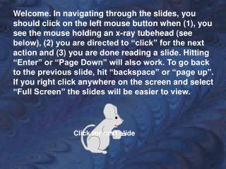
0 Welcome. In navigating through the slides, you should click on the left mouse button when (1), you see the mouse holding an x-ray tubehead (see below), (2) you are directed to “click” for the next action and (3) you are done reading a slide. Hitting “Enter” or “Page Down” will also work. To go back to the previous slide, hit “backspace” or “page up”. If you right click anywhere on the screen and select “Full Screen” the slides will be easier to view. Click for next slide
Panoramic Anatomy The following is a PowerPoint presentation. If you right click on the screen and select “Full Screen”, the images should fill the entire screen. If you want to print slides 7, 8 and 9, you must right-click, select “End Show” and then right click again on slide you want to print and select “Print”.
Types of Panoramic Images Single Real Image Double Real Image Ghost Image
Single Real Image Only one image results from a given anatomical structure. The structure is located between the rotation center and the film and the x-ray beam only passes through the structure one time. Most images seen on a panoramic film are of this type.
Double Real Image Two images of a single object are seen on the film. Double real images are produced by structures located in the midline. The x-ray beam passes through these objects twice as the tubehead rotates around the patient. Structures that result in double real images are the hard and soft palates, the hyoid bone and the cervical spine.
Ghost Image Ghost images are formed by dense objects located between the tubehead and the rotation center. These ghost images usually result from external objects such as earrings, but they may be produced by dense anatomical structures such as the mandible. (For more information, see self-study module “Panoramic Technique”). ghost image of earring (between lines)
The following slides show anatomical structures seen on panoramic films. The accompanying keys identify the structures by number. See what other structures you can identify that are not labeled. At the end of this presentation there are 11 pre-test slides.
9 12 7 19 5 17 13 25 14 6 22 18 39 28 33 9 19 7 12 17 13 14 5 6 25 22 18 39 28 33
11 2 15 24 26 32 8 23 16 1 31 3 20 4 34 44 30 36 38 2 11 15 24 32 26 8 23 16 1 31 3 20 44 34 30 36 38
46 21 41 42 47 40 45 43 46 21 41 42 47 40 45 43
R L 11 7 1 46 41 47 43 36 45 38
16 R L 17 23 2 8 6 21 18 19 39 Red arrows point to ghost image of hard palate
0 R L 11 9 3 20 • How old is this patient? • 6-9 years • 10-12 years • 13-15 years b. 10-12 years old
R L 17 2 44 20 28 43
R L 2 atlas 31 transverse foramen
0 R L 15 46 47 19 6 27 34 What head positioning error is seen on this film? The anterior teeth are positioned in front of the notch in the bitestick, resulting in the widening of the anterior teeth (the maxillary central incisors are as wide as the molars).
0 R L 17 1 8 15 32 N N = soft tissue of nose What head positioning error is seen on this film? The head is tipped down too much, resulting in shortened mandibular incisors and a V-shaped mandible.
R L 40 27 E LN 36 LN = calcified lymph node E = epiglottis
R L 0 2 8 40 18 45 ? ? Identifies calcification, possibly in carotid or in lymph node What positioning error is seen on this film? The head was turned to the left, bringing that side closer to the film and decreasing the width of the ramus on that side. The green arrow points to the biteblock, centered on the contact between the right central and lateral incisors. The patient’s head is turned to the side. Note the width of the ramus on each side (The red arrows are the same length). Which direction was the patient’s head turned (left or right)?
0 R L 8 7 46 47 33 E E = epiglottis
0 R L 11 21 3 29 32 34 What causes the black dots identifed by the red arrow? The chin is tipped up too much, giving a more squared off appearance to the mandible, creating a reverse smile and causing the hard palate to be superimposed on the roots of the maxillary teeth. The black dots result from static electricity, caused by removing the film too quickly from the cassette or from the box of film (creates friction, which results in a static discharge). What positioning error is seen on this film?
L R 16 0 10 9 20 3 42 27 30 1 44 G 36 G = ghost of right mandible
L R 0 24 14 27 47 nose 39 What caused the white (radiopaque) area indicated by the red arrow? The lead apron was placed too high on the back of the patient’s neck.
0 R L 12 air cell 9 23 7 26 Air cell in zygomatic arch.
R L 24 7 26 22 27 30 38
R L 5 10 6 47 45 ghost of mandible
R L 15 23 9 7 5 21 44 39 30 Note the relatively inferior location of the mandibular canal (30), providing plenty of room for the implant.
R L 24 26 31 1 29 Pattern on right side of film (patient’s left) caused by excessive oil on patient’s hair.
R L 7 28 28 red arrow identifies fracture
R L 27 44 34 Green arrow identifies “pseudo-fracture” caused by palatoglossal air space. Red arrows point to odontogenic keratocyst.
Ghost images of earrings L R 15 2
R L 27 28 28 Hearing aid (red arrow) with ghost (green arrow).
Ghost image of metal used to restore left angle of mandible R L
R L Ghost images of mandibles (dotted line outlines ghost of left ramus-angle over right side of mandible)
Slide # 1 0 R L C E D G F B A A B C D Cervical vertebra E F G Zygomaticotemporal suture External oblique ridge Lingula Zygomatic process Cervical vertebra Maxillary sinus
B Slide # 2 0 R L K D J E I A H F C G Hyoid bone G H I J K Ear lobe A B C D E F Mandibular canal External auditory meatus Pterygoid plates Submandibular gland fossa Articular eminence Nasal septum Pterygomaxillary fissure Hard palate Mental foramen
Slide # 3 0 L R C B D A E Palatoglossal air space A B C D E Middle cranial fossa Lateral border of the orbit Condyle Mental fossa
Slide # 4 0 L R I D E H C B A G F J K L Hard palate G H I J K L Cervical vertebra A B C D E F Post. wall of maxillary sinus Zygomaticotemporal suture External auditory meatus Zygomatic process Posterior pharyngeal wall Nasal septum Mental foramen Inferior concha Mental fossa Soft tissue of nose
Slide # 5 R L E F G C D J H B I A 0 Infraorbital canal F G H I J A B C D E Glossopharyngeal air space Infraorbital foramen Styloid process Soft palate Nasopharyngeal air space Mandibular canal Pterygoid plates Lingula Condyle
Slide # 6 0 L R E C D E B F G A E F G Pterygoid plates A B C D Mental foramen Ear lobe Incisive foramen Hyoid bone Soft tissue of nose Anterior nasal spine The radiolucency (red arrows) seen in the ramus and third molar area on the patient’s right side is an ameloblastoma. (Differential includes dentigerous cyst, radicular cyst, OKC).
Slide # 7 0 L R A B C D Posterior border of maxillary sinus A B C D Inferior border of orbit Inferior concha Inferior border of maxillary sinus The radiolucency (red arrows) seen in the ramus on the patient’s left side is a squamous cell carcinoma.
Slide # 8 L R D C A B E 0 D E Floor of middle cranial fossa A B C Maxillary tuberosity Posterior pharyngeal wall Hard palate Coronoid process This child has a condition known as cherubism. The mandibular lesions involve both rami, extending into the coronoid process (the condyle is rarely involved). The maxillary lesions are located in the tuberosity regions, causing anterior displacement of 2nd and 3rd molars.
Slide # 9 0 L R E D A C F B D E F Soft palate A B C Zygomatic arch Pterygomaxillary fissure External oblique ridge Styloid process Palatoglossal air space This patient has multiple supernumerary premolars in the mandible (#’s 21, 28 and 29 were extracted).
Slide # 10 L R C D E B A F 0 D E F Hard palate A B C Mandibular canal Mandibular foramen Soft tissue of nose Styloid process Nasal fossa This patient has impacted mandibular third molars that have migrated up into the coronoid processes. Note also the long, thin condylar necks and small condyles.
Slide # 11 0 L R B C A D E D E Articular eminence A B C Sigmoid notch Mental foramen (on crest of ridge) Nasal septum Coronoid process The green arrows identify a calcified stylohyoid ligament. If there is associated neck pain, the condition is known as Eagle’s Syndrome (recent history of neck trauma or surgery) or Stylohyoid Syndrome (no history of trauma/surgery). The red box outlines several radiopacities which represent tonsillar calcifications.