Download
1 / 24
240 likes | 407 Views
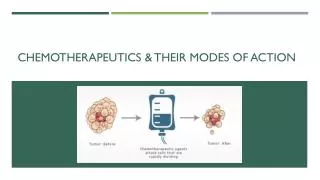
Chemotherapeutics & Their Modes of Action. Chemotherapy. Chemotherapy is the treatment of cancer with one or more cytotoxic antineoplastic drugs ("chemotherapeutic agents") as part of a standardized regimen.

E N D
Chemotherapy • Chemotherapy is the treatment of cancer with one or more cytotoxic antineoplastic drugs ("chemotherapeutic agents") as part of a standardized regimen. • Chemotherapy may be given with a curative intent or it may aim to prolong life or to palliate symptoms. • It is often used in conjunction with other cancer treatments, such as radiation therapy or surgery.
Chemotherapy • Traditional chemotherapeutic agents act by killing cells that divide rapidly, one of the main properties of most cancer cells. • This means that chemotherapy also harms cells that divide rapidly under normal circumstances: • cells in the bone marrow • digestive tract • hair follicles. • This results in the most common side-effects of chemotherapy: • myelosuppression • mucositis (inflammation of the lining of the digestive tract) • alopecia
Types of Chemotherapy • Chemotherapy drugs can be divided into several groups based on factors such as: • how they work • their chemical structure • their relationship to another drug • Because some drugs act in more than one way, they may belong to more than one group. • Knowing how the drug works is important in predicting side effects.
Types of Chemotherapy • The majority of chemotherapeutic drugs can be divided into: • Alkylating agents • Antimetabolites • Anthracyclines • Plant alkaloids • Some newer agents do not directly interfere with DNA • Monoclonal antibodies • Tyrosine kinase inhibitors, which directly target a molecular abnormality in certain types of cancer • In addition, some drugs that modulate tumor cell behavior without directly attacking those cells may be used. • Example: Hormone treatments • All of these drugs affect cell division or DNA synthesis and function in some way.
Alkylating agents directly damage DNA to prevent the cancer cell from reproducing. Target Cancers Common Side Effects Because these drugs damage DNA, they can cause long-term damage to the bone marrow. In rare cases, this can eventually lead to acute leukemia. The risk of leukemia from alkylating agents is “dose-dependent,” The risk of leukemia after getting alkylating agents is highest about 5 to 10 years after treatment. • Alkylating agents are used to treat many different cancers, including: • Leukemia • Lymphoma • Hodgkins disease • multiple myeloma • Sarcoma • cancers of the lung, breast, and ovary The platinum drugs (cisplatin, carboplatin, and oxalaplatin) are sometimes grouped with alkylating agents because they kill cells in a similar way. These drugs are less likely than the alkylating agents to cause leukemia later on.
Antimetabolitesinterfere with DNA and RNA growth by substituting for Nucleotides. Target Cancers Common Side Effects Nausea Alopecia Stomatitis Severe myelosuppression • Leukemias • Breast cancer • Ovarian cancer • Cancers of the intestinal tract
Anthracyclinesare anti-tumor antibiotics that interfere with enzymes involved in DNA replication Target Cancers Common Side Effects A major consideration when giving these drugs is that they can permanently damage the heart if given in high doses. For this reason, lifetime dose limits are often placed on these drugs. • Prostate cancer • Breast cancer • Lymphoma • Leukemia
Topoisomerase inhibitors Target Cancers Common Side Effects Treatment with topoisomerase II inhibitors increases the risk of a second cancer — acute myelogenous leukemia (AML). With this type of drug, a secondary leukemia can be seen as early as 2 to 3 years after the drug is given. • Certain leukemias • Lung cancer • Ovarian cancer • Gastrointestinal cancer • Other cancers
Mitotic inhibitors are often plant alkaloids and can stop mitosis or inhibit enzymes from making proteins needed for cell reproduction. Target Cancers Common Side Effects Known for their potential to cause peripheral nerve damage, which is a dose-limiting side effect. • Breast cancer • Lung cancer • Myelomas • Lymphomas • Leukemias
Paclitaxel • Paclitaxel is a mitotic inhibitor used in cancer chemotherapy to treat patients with • Lung cancer • Ovarian cancer • Breastcancer • Head and neck cancer • Advanced forms of Kaposi's sarcom • It was discovered in1967 after being isolated it from the bark of the Pacific yew tree, Taxusbrevifolia • it was named taxol.
TAXOL • 1962--as part of the exploratory program bark from the pacific Yew was collected by USDA. It showed activity in an initial in vitro screen. • 1971--Dr. Monroe Wall reports the isolation and identification of taxol. • 1972--Shows only moderate activity against murine tumor cell lines • 1975--Shows strong activity against melanoma • 1977--Accepted as a drug candidate for NCI preclinical development • 1980--toxicology studies begin • 1983--Phase I clinical trial begins (cremophor) • Supply dwindling (1 tree/treatment) • Taxol is now synthesized by semi-synthetic route using leaves from tree
TAXOL: Lessons learned • Use human tumor cells for screening instead of murine cells • Formulation issues need to be addressed early on • Mechanism of action is important in drug development process • Critical need to address supply issue at an early stage of development
Paclitaxel Mode of Action • Paclitaxel stabilizes microtubules and as a result, interferes with the normal breakdown of microtubules during cell division.
Roles of Microtubules in the cell • The cytoskeleton is a network of fibers extending throughout the cytoplasm • It organizes the cell’s structures and activities, anchoring many organelles • It is composed of three types of molecular structures • Microtubules • Microfilaments • Intermediate filaments
Microtubules • Microtubules are hollow rods about 25 nm in diameter and about 200 nm to 25 microns long • Functions of microtubules • Shaping the cell • Guiding movement of organelles • Separating chromosomes during cell division
Microtubule Formation • In the cell itself, microtubules are formed in an area near the nucleus called the "aster". • This is also called the Microtubule Organizing Center (MTOC). • The first stage of formation is called "nucleation". • The process requires tubulin, Mg++ and GTP. • This stage is relatively slow until the microtubule is initially formed. • During "nucleation", an alpha and a beta tubulin molecule join to form a heterodimer. • In the second phase called “elongation” these attach to other dimers to form oligomers which elongate to form protofilaments. • http://sites.sinauer.com/cooper5e/animation1203.html Usually the minus end is the anchor point in the MTOC Microtubules are polar with a plus end (fast growing) and a minus end (slow growing). + -
Cilia and Flagella • Microtubules control the beating of cilia and flagella, locomotor appendages of some cells • Cilia and flagella share a common structure • A core of microtubules sheathed by the plasma membrane • A basal body that anchors the cilium or flagellum • A motor protein called dynein, which drives the bending movements of a cilium or flagellum
Centrosomes and Centrioles • In many cells, microtubules grow out from a centrosome near the nucleus • The centrosome is a “microtubule-organizing center” • In animal cells, the centrosome has a pair of centrioles, each with nine triplets of microtubules arranged in a ring that are “thought” to be involved with cell division
Polymerization and depolymerization microtubule dynamics • Microtubules consisting of α/β heterodimers elongate to form cylindrical microtubules of 13 protofilaments with a plus (+) end and minus (−) end. • Tubulin-bound GTP binds to plus (+) end of microtubule and GTP is hydrolyzed to GDP + Pi forming a GTP cap. • The GTP cap stabilizes the microtubules plus (+) end and stabilization is further enhanced by addition of the taxanes, paclitaxel and docetaxel, which bind to β-tubulin sites causing polymerization of microtubules. • Depolymerizationoccurs when the tubulin − GTP / GDP + Pi cap is lost, Pi is released from tubulin, destabilization occurs and the tubulin-bound GDP dissociates from the plus (+) end causing depolymerization
Vesicle ATP Figure 6.21 Receptor formotor protein Microtubuleof cytoskeleton Motor protein(ATP powered) (a) Microtubule Vesicles 0.25 m
Microtubules are dynamic • Dynamic instability refers to the coexistence of assembly and disassembly at the (+) end of a microtubule. • The microtubule can dynamically switch between growing and shrinking phases in this region. • This constant growing and shrinking is necessary for microtubules to carry out their functions • During polymerization, both the α- and β-subunits of the tubulin dimer are bound to a molecule of GTP • The GTP bound to α-tubulin is stable and it plays a structural function in this bound state. • GTP bound to β-tubulin may be hydrolyzed to GDP shortly after assembly resulting in the addition of new dimer
The kinetics of GDP-tubulin are different from those of GTP-tubulin • GDP-tubulin is prone to depolymerization • Tubulin adds onto the end of the microtubule only in the GTP-bound state • Acap of GTP-bound tubulin at the tip of the microtubule can protect it from disassembly. • When hydrolysis catches up to the tip of the microtubule, it begins a rapid depolymerization and shrinkage
Paclitaxel binds to the interior surface of the microtubule at the taxane-binding site, suppressing microtubule dynamics • Paclitaxel has a specific binding site on the microtubule polymer, and has the ability to polymerize tubulin in the absence of cofactors like guanosine triphosphate and microtubule-associated proteins