Understanding the Cell Cycle: From Interphase to Cytokinesis
100 likes | 216 Views
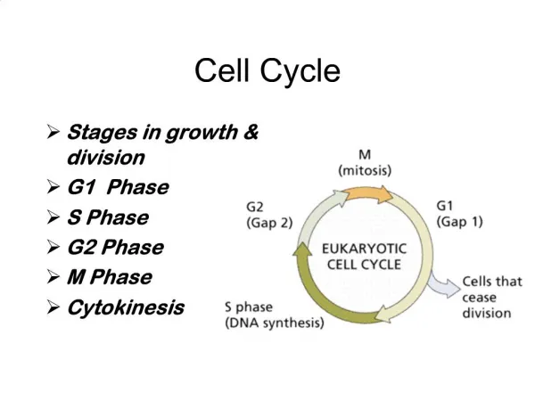
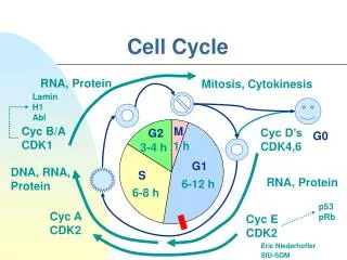
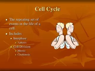
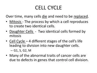
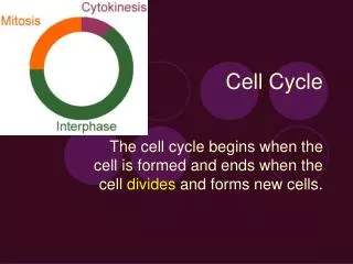
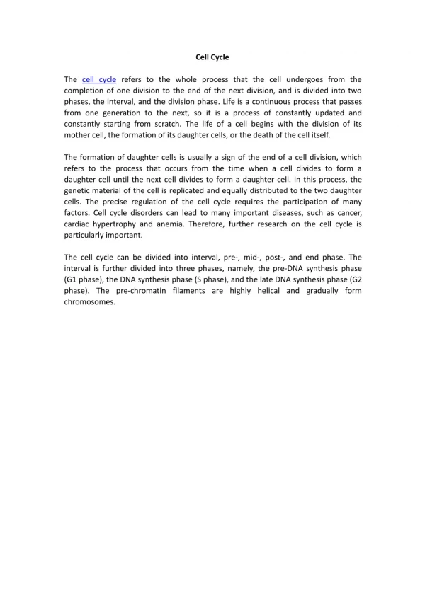
This informative guide explores the cell cycle stages, including interphase, mitosis, and cytokinesis. Interphase is the longest phase where cells spend the majority of their time preparing for division. Mitosis consists of prophase, prometaphase, metaphase, anaphase, and telophase, each playing a crucial role in the separation of DNA. Finally, cytokinesis divides the cytoplasm, resulting in two daughter cells. Each phase is illustrated with detailed figures and links to additional resources for deeper understanding.

Understanding the Cell Cycle: From Interphase to Cytokinesis
E N D
Presentation Transcript
Cell cycle By: Caleb Calhoun
interphase Cells are in this phase most of the time. Figure 1 Figure 2 fig 1 http://emt.bu.edu/em610/em610_ol_spring_2008/yanti/interphase.html Fig 2 http://micro.magnet.fsu.edu/cells/fluorescencemitosis/interphase1large.html
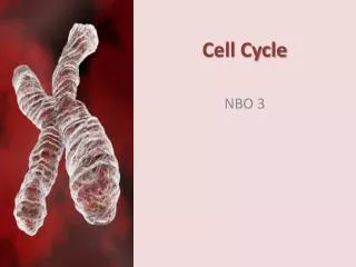
mitosis • Prophase • Prometaphase • Metaphase • Anaphase • Telophase
Mitosis: prophase Spindle fibers appear. Figure 1 Figure 2 Fig 2 http://www.prism.gatech.edu/~gh19/b1510/mitmei.htm Fig 1 http://itg.beckman.illinois.edu/technology_development/web_atlas/structures/mitosis/prophase/
prometaphase Prometaphase is when the DNA finds it’s match and connects together. Figure 1 Figure 2 Fig 1 http://www.sciencephoto.com/media/214742/enlarge Fig 2 http://www.edupic.net/cells.htm
metaphase Metaphase is when the DNA lines up in the middle of the cell and the spindle fibers hold them. Figure 1 Figure 2 Fig 2 http://en.wikipedia.org/wiki/File:Metaphase.png fig 1. http://faculty.washington.edu/casbury/
anaphase The spindle fibers pull the DNA two them. Figure 1 Figure 2 Fig 1 http://upload.wikimedia.org/wikipedia/commons/thumb/9/91/Anaphase_IF.jpg/300px-Anaphase_IF.jpg Fig 2 http://www.yvonnebraden.com/Mitosis%20Flip%20Book%20Warm-up.htm
Telophase The cell membrane or cell wall appears and splits the cell. Figure 2 Figure 1 Fig 1 http://micro.magnet.fsu.edu/cells/fluorescencemitosis/telophase1large.html Fig 2 http://en.wikipedia.org/wiki/File:Telophase.jpg
Cytokinesis Cytokinesis is when the two cells a finally apart. Figure 1 Figure 2 Fig 1 http://images.tutorvista.com/content/feed/tvcs/cytokin.gif Fig 2 http://team6cell.pbworks.com/f/telophase.gif