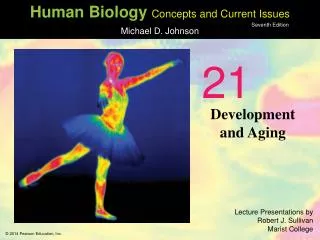
Development and Aging
Development and Aging. 0. 21. Fertilization Begins When a Sperm and Egg Unite. Sperm and egg unite 6–24 hours after intercourse Fertilization occurs in the upper third of the oviduct One sperm fertilizes one egg, forming a zygote Typical ejaculate: several hundred million sperm

Development and Aging
E N D
Presentation Transcript
Developmentand Aging 0 21
Fertilization Begins When a Sperm and Egg Unite Sperm and egg unite 6–24 hours after intercourse Fertilization occurs in the upper third of the oviduct One sperm fertilizes one egg, forming a zygote Typical ejaculate: several hundred million sperm Only a few hundred to a thousand sperm make it to the upper oviduct Many barriers and challenges on the sperm’s journey Distance Vaginal pH Cervical mucus White blood cells
One Sperm Fertilizes the Egg Several hundred to a thousand sperm reach the upper oviduct, but only one fertilizes the egg Fertilization Acrosome—in head of sperm Digestive enzymes which penetrate the egg Sperm proteins lock with egg membrane receptors Zona pellucida becomes impermeable to other sperm Sperm actually penetrates the secondary oocyte, which then completes meiosis, forming haploid ovum Fertilization occurs when sperm nucleus fuses with ovum nucleus, forming diploid zygote
Figure 21.1 Optimal siteof fertilization Oviduct Uterus Secondaryoocyte Acrosome Head Coronaradiata Sperm Ovary Midpiece Cytoplasm Tail Nucleus Plasmamembrane Cervix First polarbody Vagina Zonapellucida The female gamete. The femalegamete is a secondary oocyte thatis in an arrested state of stage II ofmeiosis. The male gamete, or sperm.The size of a sperm relative tothe secondary oocyte has beengreatly exaggerated. Fertilization. Fertilizationgenerally takes place in theupper third of the oviduct.
Figure 21.2 Sperm Corona radiata Secondary oocyte First polar body Sperm nucleus Acrosome All otherspermdeniedentrance Acrosomereleases enzymes Granulosa cellsof corona radiata Zona pellucida Oocyte and spermplasma membranes fuse Oocyte plasmamembrane Granules releaseenzymes that make thezona pellucida impenetrable Granule Oocytecytoplasm Sperm nucleus engulfedby oocyte cytoplasm
Twins May Be Fraternal or Identical Fraternal twins Ovulation of more than one oocyte, each of which is fertilized by different sperm No more similar than two siblings May be different genders Identical twins One oocyte fertilized Split into two pre-embryos before 16-cell stage Same gender, look alike
Figure 21.4 Identical twins arisewhen a zygote divides intwo during development. Fraternal twins arisewhen two eggs areovulated and fertilized inthe same monthly cycle.
Development: Cleavage, Morphogenesis, Differentiation, and Growth Cleavage Series of cell divisions without cell growth Produces a ball of identical cells Occurs up to about day four after fertilization Occurs entirely within the oviduct Morphogenesis Changes in shape and form Physical development of organism Ongoing At implantation, growth in size
Development: Cleavage, Morphogenesis, Differentiation, and Growth Differentiation Individual cells take on specialized forms and functions Growth Begins significantly at implantation Single cell at fertilization to trillion cells at birth Growth in number of cells and size of cells results in increase in overall size
Pre-Embryonic Development: The First Two Weeks Conceptus travels through oviduct Growth, differentiation, and morphogenesis begin Morula (ball of 32 identical cells)—results from cleavage Blastocyst (hollow ball with inner cell mass that will become the embryo)—results from differentiation Implantation occurs as the blastocyst burrows into the endometrium day six or seven post-fertilization Embyronic disk (destined to become the embryo) develops—end of pre-embryonic period
Pre-Embryonic Development Ectopic pregnancy Occurs when blastocyst implants in an oviduct prior to reaching the uterus Oviduct is not large enough to support the development of a full-term baby Ectopic pregnancies are often terminated to protect the health of the mother
Figure 21.5 Zygote Diploidnucleus Zonapellucida Uterinetube Morula Ovary Zona pellucida regresses Blastocyst Inner cellmass Hollow cavity Trophoblast
Figure 21.6 Amniotic cavity Ectoderm Cells derivedfrom trophoblast Endometrium Endoderm Innercellmass Amnioticcavity Yolk sac Embryonicdisk Trophoblast Blastocyst
Embryonic Development: Weeks Three to Eight Rapid growth, differentiation, morphogenesis All organs, organ systems established by end of embryonic period Embryonic development: beginning of week three until end of week eight
Tissues and Organs Derive from Three Germ Layers Ectoderm: outermost layer Becomes epidermis, nervous system, hair, nails, tooth enamel, parts of eye Mesoderm: middle layer Becomes muscle, connective tissue, bone, kidneys, ureters, testes, ovaries Endoderm: innermost layer Becomes liver, pancreas, alveoli, bladder lining, lining of urethra, vagina
Figure 21.7 Amnioticcavity Trophoblastcell Ectoderm Mesoderm Embryonicdisk Endoderm Endometrialcells Yolk sac
Extra-Embryonic Membranes Develop early in embryonic development Amnion Lines amniotic cavity, filled with amniotic fluid Cushions the fetus Allantois Temporary, helps form blood vessels of umbilical cord Yolk sac Source of assorted cells for organ development Chorion Makes human chorionic gonadotropin (hCG), which supports pregnancy for the first three months Forms structures that will become part of placenta
The Placenta and Umbilical Cord Development Chorion digests into endometrium, creating pool of blood Placenta seals off the pool and projects chorionic villi into maternal blood Villi contain blood capillaries connected through the umbilical vessels to the fetus Umbilical cord: two-way life line, connects placenta to embryo’s circulation
The Placenta and Umbilical Cord Functions Filters nutrients, waste, and antibodies for the fetus without mixing mother and fetal circulations Site of nutrient and gas exchange between embryo and mother Some toxins or viruses may pass through Alcohol, cocaine, HIV Endocrine Initially produces hCG (human chorionic gonadotropin Later produces estrogen and progesterone
Figure 21.8 Fetal portionof placenta(chorion) Maternal portionof placenta(endometrium) Placenta Yolk sac Amniotic cavitycontainingamniotic fluid Fetal arteriole Fetal venule Umbilicalcord Amnion Uterus Umbilical cord Amnion Connectionto yolk sac Cavity of uterus Maternalvein Maternalartery Umbilicalarteries Umbilicalvein Pool ofmaternalblood Chorionicvilluscontainingfetal capillaries A closer view of portions of the placenta and umbilical cord,showing how nutrients and gases are exchanged betweenmaternal and fetal blood without mixing. The fetus is bathed in amniotic fluid withinthe amnion. Its only connection to the motheris the umbilical cord.
The Embryo Develops Rapidly Day 15 Embryonic disk elongates along one axis Primitive streak appears in embryonic disk Days 19–24 Neural tube develops: becomes brain and spinal cord Pharyngeal arches develop Somites (segments): bone, muscle, skin End of week four Heart is beginning to develop Eye development begins Limb buds appear
Figure 21.9 Primitivestreak Pharyngealarches Neuralgroove Future brain Somites Day 14 Day 15 Days 19–23 Day 25
Figure 21.10 Week 4 Yolk sac Actual length Embryo Connecting stalk Forebrain Future lens Pharyngeal arches Developing heart Upper limb bud Somites Neural tube forming Lower limb bud Tail
Animation: Embryonic Development Right-click and select Play
Gender Development Begins at Six Weeks Y chromosome gene for sex-determining region Y (SRY) activated Testes-determining factor synthesized from SRY gene—will initiate development of the testes Testes secrete two hormones 1. Testosterone stimulates the further development of male genitalia 2. Anti-Mullerian hormone suppresses development of female external and internal genitalia Absence of Y chromosome results in female development
Figure 21.12 Bud Labioscrotal swelling Urogenital groove Y chromosomeabsent Y chromosomepresent 6 weeks 10 weeks Development does not requirehormones. Development requirestestosterone fromdeveloping testes. Vaginalopening Testes Birth approaching
Months Three and Four Eight weeks: marks the transition from embryonic to fetal development Months three and four Organ development Beginnings of organ function Kidneys, liver, spleen Cartilaginous skeleton replaced with bone Bone marrow begins producing red blood cells Face develops Rapid growth
Months Five and Six Months five and six Fetal movement begins Fetal heartbeat can be heard with stethoscope Fetus responds to external sounds Lungs produce surfactant—significant for survival outside of mother Survival possible outside mother End of six months marks end of second trimester
Months Seven through Nine Months seven through nine (third trimester) Rapid growth and maturation Fetal activity increases Fetus prepares for life “on the outside” Lungs and digestive tract ready to function Average size at birth 20 inches in length, 6–7.5 lbs.
Birth and the Early Postnatal Period Labor initiated by hormones secreted by maturing fetus Uterus begins to contract and contractions strengthen with continuous positive feedback Stages of labor Stage 1: dilation of cervix Stage 2: expulsion of fetus (delivery) Stage 3: afterbirth—expulsion of placenta Cesarean delivery Surgical delivery of baby
Figure 21.14 Placenta Urinary bladder Rupturedamnion Pubic bone Urethra Vagina Cervix Rectum 9-month-old fetus Stage 1: dilation. The cervical opening widens.The amnion may break at this stage. 9-month-old fetus. As birth approaches,the fetus usually is positioned with the headdown and toward the cervix. Placenta Uterus Umbilicalcord Stage 3: afterbirth. The placenta detachesfrom the uterus and is expelled along with theremainder of the umbilical cord. Stage 2: expulsion. The fetus passes headfirstthrough the cervical canal and the vagina.
The Transition from Fetus to Newborn Taking the first breath Pulmonary surfactants necessary for alveoli to expand and fill with air Changes in cardiovascular system Umbilical circulation cut off Ductus venosus regresses to connective tissue Foramen ovale and ductus arteriosus close in days/weeks, cause blood to go through the lungs instead of bypassing them All blood from the digestive tract goes to liver
Figure 21.15 Moderate oxygenation Low oxygenation Very low oxygenation High oxygenation Aorta Pulmonaryartery Ductus arteriosus(becomes connectivetissue) Superiorvena cava Left atrium Leftventricle Right atrium Foramen ovale(closes shortly afterbirth) Right ventricle Lung Inferiorvena cava Hepaticportal vein(fromdigestiveorgans) Ductus venosus(becomes connectivetissue) Liver Abdominalaorta Inferiorvena cava Umbilical vein(becomes connectivetissue) Umbilical cord Commoniliac artery Umbilical arteries(become connectivetissue) Fetal circulation. Circulation after birth.
Lactation Produces Milk to Nourish the Newborn Endocrine control of lactation Estrogen and progesterone cause breast enlargement during pregnancy Prolactin stimulates milk production Oxytocin stimulates smooth muscle contractions that cause ejection of milk Colostrum Watery milk produced the first few days after birth Rich in antibodies
From Birth to Adulthood Neonatal period (first month) Helpless period—movement by reflex rather than by conscious control Infancy (2 to 15 months) Rapid development and maturation of organ systems Rapid brain growth, particularly cerebral cortex Childhood (16 months to 12 years) Continued development and growth Adolescence (15 to 20 years) Growth spurt Sexual maturation
Figure 21.16 3months 2years 5years 13years newborn 22years 80years 2months
Aging Takes Place Over Time Aging: process of change associated with the passage of time Senescence: the progressive deterioration of organs and multiple organ systems over time Longevity: how long a person lives Though life spans increased throughout the 1900s, this was due to reduction in deaths by accident and disease, NOT due to slowing of the aging process
What Causes Aging? Three hypotheses 1. Internal genetically determined program that counts finite number of cell divisions, thus determining cell death 2. Accumulation of cell damage or errors limits cells’ ability to repair themselves Damage accelerated by oxygen free radicals 3. Aging is a whole-body process; all systems are interdependent and the decline in function of a critical body system may lead to parallel senescence of other systems
Figure 21.18 Useful genesection of DNA Telomere sectionof DNA Nucleotideslost fromtelomere Old (template)strand Newly formingstrand
Body Systems Age at Different Rates Bone and muscle mass decrease in early adulthood Bone mass loss accelerates in women after menopause Skin becomes thinner, less elastic Lung tissue decreases in elasticity, decreasing lung capacity Blood vessels lose elasticity, heart walls become stiffer and less efficient Blood pressure may increase
Body Systems Age at Different Rates Thymus decreases in size/function—fewer T cells, declining immune response Brain and sensory functions decline, affecting every neural activity Hearing loss due to stiffening of hair cells within the cochlea Vision problems due to stiffening of lens Female reproductive capability ceases at menopause Male reproductive capability declines over time
Body Systems Age at Different Rates Decreased ability of the liver to detoxify and metabolize drugs Kidney mass decreases, filtration rate decreases, but generally of little consequence Many men experience prostate hypertrophy Women may experience urinary incontinence
Aging Well Lifestyle Exercise Diet
Death Is the Final Transition Failure of critical organ systems leads to rapid death Brain Respiratory system Cardiovascular system Death is a process; defining the moment of death is a challenge Legal and medical criteria of death Irreversible cessation of circulatory and respiratory functions Irreversible cessation of all functions of the entire brain, including the brain stem