Chapter 12- CNS and epidermis
130 likes | 469 Views
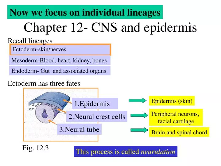
Now we focus on individual lineages. Chapter 12- CNS and epidermis. Recall lineages. Ectoderm-skin/nerves. Mesoderm-Blood, heart, kidney, bones. Endoderm- Gut and associated organs. Ectoderm has three fates. Epidermis (skin). 1.Epidermis. Peripheral neurons, facial cartilage.

Chapter 12- CNS and epidermis
E N D
Presentation Transcript
Now we focus on individual lineages Chapter 12- CNS and epidermis Recall lineages Ectoderm-skin/nerves Mesoderm-Blood, heart, kidney, bones Endoderm- Gut and associated organs Ectoderm has three fates Epidermis (skin) 1.Epidermis Peripheral neurons, facial cartilage 2.Neural crest cells 3.Neural tube Brain and spinal chord Fig. 12.3 This process is called neurulation
Neural plate Dorsal ectoderm becomes neural ectoderm to become neural plate to become neural tube Primary Neurulation Neural crest 1. Folding epidermis • Two types of neurulation • Primary- “pinching off” • Secondary – hollow out a cord • Both are used in many creatures 2. elevation 3. convergence 4. closure Fig. 12.4- Amphibian embryo Neural tube Fig. 12.3
A few details at each step in primary neurulation 1. Folding Mesoderm signals ectodermal cells to form neural plate 2. Elevation and 3. Convergence Mesoderm signals ectodermal cells to form neural plate Hinge cells (called medial hinge point cells) attached to notochord Cell shape and cells movement contribute to elevation Fig. 12.6 4. Closure Folds adhere to each other Failure of complete closure results in neural tube defects • anacephaly – anterior tube fails to close- • brain development ceases • Spina bifida – posterior tube fails to • close at human day 27 • 50% of spina bifida preventable with 0.4mg/day vitamin B12
Secondary neurulation A cord is first made, then hollowed out Example- posterior end of chick Note- rest of chick uses primary neurulation Further neural tube differentiation 1. Anterior-posterior axis Anterior portion of neural tube forms three vesicles: 1. Forebrain 2. Midbrain 3. Hindbrain Brain volume increases 30-fold between days 3 and 5 of development
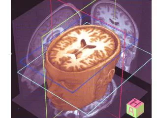
Brain development is complex and laden with nomenclature Fig. 12.10- human brain development
2. Dorsal-ventral axis Fig. 12.13- chick neural tube Epidermis (then roof plate) secretes TGF-b family proteins (BMP-4 and –7, dorsalin, activin) to signal dorsal portion of neural tube to become sensory neurons • Notochord (then hinge cells) secretes sonic hedgehog to signal ventral portion of neural tube to become motor neurons • Retinoic acid also plays a role Roof plate Hinge cells
Neuronal types • Brains consists of 1011neurons (nerve cells) and 1012glia (support cells) • The long-held belief that neurons were fully determined at birth is incorrect- • Evidence for neuronal stem cells exists Cells lining neural tube can give rise to neurons or glia cells Fig. 12.22- A motor neuron Input axons from other neurons Growth cone Axon • At birth, very few dendrites are present on cortical neurons • Cortical neurons connect to 10,000 other neural cells during 1st year post birth!! Dendrites- connect to other neurons • Axons are part of the cell body that can extend several feet • Growth cone exploresand moves into new regions of body
Nerve cells are protected to facilitate electrical signal conduction by: In central nervous system In peripheral nervous system By myelination from Schwann cells By myelin sheath produced by oligodendrocytes Pax gene expression Vertebrate eye development Pax6 gene encodes protein that directs eye development Neural-tube specific enhancer Fig. 5.15-the Pax 6 gene
Recall chapter 5- introduce DNA containing pax6 cDNA under control of an inducible promoter + a tissue-specific enhancer Fig. 5.14 Observe additional eyes Pax6 mutants lack eyes in flies, mice and humans • Sonic hedgehog dictates formation of two eyes • Mutants produce one eye (cyclopia) Fig. 6.25- a cyclopic lamb
Eye development requires the specification of numerous tissues Eye lens development forms by: 1. Lens vessicle folds onto itself to form ring Fig. 12.29 2. Interior cells elongate across cavity to produce crystallin lens fibers 3. Cells enucleate Fig. 12.27
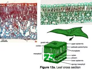
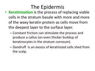
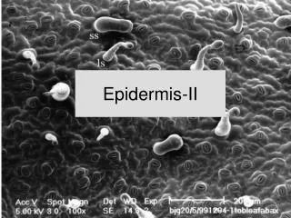
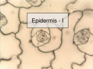
A few words about epidermis (skin) development Recall: Epidermis becomes two layers, a periderm (which is shed) and a basal layer that gives rise to skin cells (Shed) Periderm Epidermis Basal layer Granular cells Keratinocytes Spinous layer Termed “Malpighian layer”
Fig. 13.32 Keratinocytes (continually shed) TGF-a and FGF7 are important factors in skin development Granular layer Spinous layer Malpighian layer Basal layer Cells differentiate and migrate toward surface Feather, hair and scales are formed by epithelial-mesenchymal interactions between epidermis and mesoderm