RECTAL PROLAPSE
RECTAL PROLAPSE. Rectal Prolapse:. Prolapse of the rectum mainly two types: Partial or incomplete prolapse (procidentia) when the mucous membrane lining the anal canal protrudes through the anus only. Complete prolapse in which the whole thickness of


RECTAL PROLAPSE
E N D
Presentation Transcript
Rectal Prolapse: Prolapse of the rectum mainly two types: Partial or incomplete prolapse (procidentia) when the mucous membrane lining the anal canal protrudes through the anus only. Complete prolapse in which the whole thickness of the bowel protudes through the anus. Rectal prolapse occurs most often at extremes of life e.g, in children between 1-5 years of age and elderly people. More common in female than male.
the predisposing causes are:- The vertical straight course of the rectum. Reduction of supporting fat in the ischiorectal fossa. Straining at stool. Chronic cough. Aetiology In children:
In adult: Partial prolapse the predisposing causes depend on type of the prolapse. Advance degree of prolapsing piles. Loss of sphincteric tone. Straining from urethral obstruction. Operations for fistula. is generally regarded as sliding hernia of the recto vesical or recto vaginal pouch due to stretching of the levator from pregnancy, obesity. Complete prolapse
Clinical Features Prolapse is first noted during defaecation. Discomfort during defaecation. Bleeding. Mucous discharge. Bowel habit irregular and may lead to incontinence.
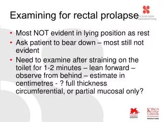
Examining for rectal prolapse • Most NOT evident in lying position as rest • Ask patient to bear down – most still not evident • Need to examine after straining on the toilet for 1-2 minutes – lean forward – observe from behind – estimate in centimetres - ? full thickness circumferential, or partial mucosal only?
Ano-rectal digital examination • Resting tone (low = IAS problem) • Squeeze pressure (low = EAS problem) • Co-ordination • Sensation (? Neurological dysfunction) • Assessment stops here for MOST patients
Complications of rectal prolapse: Irreducibility (table sugar!) Infection Ulceration Severe haemorrhage from one of the mucosal vein Thrombosis and obstruction of the venous returns leading to oedema Irreducibility and gangrene
TREATMENT Prolapse in children: the prolapse tends to disappear spontaneously by the age of 5 years. So conservative measures are sufficient. Conservative treatment: constipation and straining at stool are avoided and the buttocks may be strapped together to discourage prolapse during defaecation. Perirectal injection of alcohol/phenol may be used to fix the lax mucosa to underlying tissue. ANORECTAL DISORDERS
Prolapsed in Adult Injections of 5% phenol in oil in submucosa. 10-15ml total. Electrical stimulation with sphincteric exercises.
Surgery always necessary, none are ideal. Thiersch’s operation Rectopexy Rectosigmoidectomy Ivalon sponge rectopexy Ripstein operation Low anterior resection (minor)
2005 Estimated US Cancer Deaths* • 15% Breast • 10% Colon and rectum • 6% Ovary • 6% Pancreas • 4% Leukemia • 3% Non-Hodgkin lymphoma • 3% Uterine corpus • 2% Multiple myeloma • 2% Brain/ONS • 22% All other sites • 27% Lung and bronchus Lung and bronchus 31% Prostate 10% Colon and rectum 10% Pancreas 5% Leukemia 4% Esophagus 4% Liver and intrahepatic 3%bile duct Non-Hodgkin 3% Lymphoma Urinary bladder 3% Kidney 3% All other sites 24%
Decreasing mortality of CRC 5-year Survival 1960-70 1980-90 Colon cancer 40-45% 60% Rectal cancer 35-40% 58%
Anatomic Location of CRC • Cecum 14 % • Ascending colon 10 % • Transverse colon 12 % • Descending colon 7 % • Sigmoid colon 25 % • Rectosigmoid junct.9 % • Rectum 23 % 70%
Epidemiology • Increasing Incidence of CRC • Incidence 30-40 / 100000 / year • >70 y. of age 300 / 100000 / year • third most common malignant disease • second most common cause of cancer death
Epidemiology • 70% of CRC are resectable at diagnosis • Mortality has decreased
Ethiology • Diet: fibers, vit E, vit C • Polips (adenomatous) • IBD – more then 10 years of progression • Smoking • Cyclooxigenase inhibitors • Genetic cancer
WHO Classification of CRC • Adenocarcinoma in situ / severe dysplasia • Adenocarcinoma • Mucinous (colloid) adenocarcinoma (>50% mucinous) • Signet ring cell carcinoma (>50% signet ring cells) • Squamous cell (epidermoid) carcinoma • Adenosquamous carcinoma • Small-cell (oat cell) carcinoma • Medullary carcinoma • Undifferentiated Carcinoma
Symptoms • Bleeding per anum • Sensation of incomplete bladder empting • Tenesmus • Abdominal pain • Palpable rectal tumor • Pacienţi în stadii avansate: pierdere ponderală, hepatomegalie, icter, anemie. • Examenul fizic include: aprecierea stării generale, a prezenţei adenopatiilor periferice şi a hepatomegaliei. !!! RECTAL EXAMINATION
Investigations • Staging: - Recto- and colonoscopy - Barium enema - CT - MRI - EUS
RECTOSCOPY • COLONOSCOPY + BIOPSY Indications • - Suggestive images on barium enema • - Suggestive symptoms of colonic cancer • - Screening • -After polipectomy
EUS • Accuracy 81-93% • More difficult to interpret • Limited value in evaluation of LN invasion • Requires contact with tumor and a lumen in which to be inserted.
Tumor markers • CEA • CA 19-9 • Dynamic may be significant for recurrence
Clinical Staging of CRC Astler-Coller modified Dukes stage TNM Primary Lymph-node Distant Dukes stage tumor metastasis metastasis stage Stage 0 Tis N0 M0 A A Stage I T1 N0 M0 A A1 T2 N0 M0 A B1 Stage II T3 N0 M0 B B2 T4 N0 M0 B B2 Stage III A any T N1 M0 C C1/C2 B any T N2, N3 M0 C C1/C2 Stage IV any T any N M1 D D
TNM Classification Tis T1 T2 T3 T4 Extension to an adjacent organ Mucosa Muscularis mucosae Submucosa Muscularis propria Subserosa Serosa
Stage and Prognosis Stage 5-year Survival (%) 0,1 Tis,T1;No;Mo > 90 I T2;No;Mo 80-85 II T3-4;No;Mo 70-75 III T2;N1-3;Mo 70-75 III T3;N1-3;Mo 50-65 III T4;N1-2;Mo 25-45 IV M1 <3
Purpose of Radio(chemo)therapy in Rectal Cancer • To lower local failure rates and improve survival in resectable cancers • to allow surgery in primarly inextirpable cancers • to facilitate a sphincter-preserving procedure • to cure patients without surgery: very small cancer or very high surgical risk
Rectal Cancer • Surgery is the mainstay of treatment of RC • After surgical resection, local failure is common • Local recurrence after conventional surgery: • 15%-45% (average of 28%) • Radiotherapy significantly reduces the number of local recurrences
Radiotherapy in the management of RC • Preoperative RT (30+Gy): 57% relative reduction of local failure • Postoperative RT (35+Gy): 33% relative reduction
ESMO Recommendations • Resectable cases • Surgical procedure: TME • Preoperative RT: recommended • Postoperative chemoradiotherapy: T3,4 or N+ • Non-resectable cases: local recurrences • Preoperative RT with or without CT
Surgery-related -Low anterior resection -Excision of the mesorectum -Extent of lymphadenectomy -postoperative anastomotic leakage -Tumor perforation Tumor-related -Anatomic location -Histologic type -Tumor grade -Pathologic stage -radial resection margin -neural, venous, lymphatic invasion Predicting risk of recurrence in RC
Total Mesorectal Excision (TME) • Local recurrence rates after surgical resection of RC have decreased from about 30% to < 10% • 1. Radio(chemo)therapy • 2. Importance of circumferential margin (TME)
Epidermoid carcinoma • 75% of all malignancies of the area • Early: verucous, nodular lesion • Late: ulcerated, indurated, nodular nmass • Palpable inguinal nodes • May invade the rectum: false impression of rectal carcinoma • Lymphatic spread: like rectal + inguinal nodes
Treatment • External radiation + concomitant chemotherapy • Radical surgery in case of failure
Malignant melanoma • Horrible prognosis • Dark mass protruding from the anus • 50% pigmented • Lymph node MTS early • Treatment - not clear advantage of any alternative
Bowen’s disease:Squamous cell carcinoma in situ • Like all other places of skin • Plaque-like eczematoid lesion + pruritus • Biopsy-carcioma in situ + hyperkeratosis and giant cells • Therapy: local excision with safety margins
Basal cell carcinoma • Ulcerating tumor (uncommon) • “Rodent ulcer” like every other place of skin exposed • Doesn’t spread distantly • Local excision
Paget’s disease • Rare condition • Pale plaquelike condition with induration + nodular mass (not always) • Nodular mass= coloid carcinoma from glands or other skin appendages • Local excision (without mass) • Radical surgery + chemo + RT for coloid carcinoma