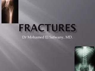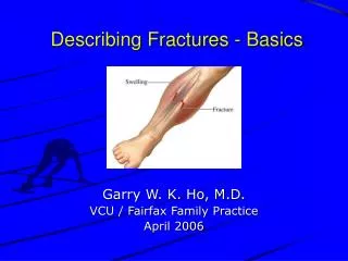
Describing Fractures - Basics
440 likes | 1.21k Views
Describing Fractures - Basics. Garry W. K. Ho, M.D. VCU / Fairfax Family Practice April 2006. Relevance. Important to know how to describe fractures Documentation Communicate with other physicians Colleagues Specialists “Ortho-speak”. Pre-reading Musculoskeletal Radiographs.

Describing Fractures - Basics
E N D
Presentation Transcript
Describing Fractures - Basics Garry W. K. Ho, M.D. VCU / Fairfax Family Practice April 2006
Relevance • Important to know how to describe fractures • Documentation • Communicate with other physicians • Colleagues • Specialists • “Ortho-speak”
Pre-reading Musculoskeletal Radiographs • 1: Name, date, old films for comparison • 2: What type of view(s) • 3: Identify bone(s) & joint(s) demonstrated • 4: Skeletal maturity (physes: growth plates) • 5: Soft tissue swelling • 6: Bones & joints (fractures & dislocations)
What is a (bony) fracture? • Disruption of a bone’s normal structure or “wholeness” • Crack, break, or rupture in a bone • There are many how’s and why’s to bony fractures • Terms used to describe each are related
Mnemonic: OLD ACID • O: Open vs. closed • L: Location • D: Degree (complete vs. incomplete) • A: Articular extension • C: Comminution / Pattern • I: Intrinsic bone quality • D: Displacement, angulation, rotation
O: Open vs. Closed • Open fracture • AKA: “Compound fracture” • A fracture in which bone penetrates through skin; • “Open to air” • Some define this as a fracture with any open wound or soft tissue laceration near the bony fracture • Closed fracture • Fracture with intact overlying skin
L: Location Epiphysis • Which bone? • Thirds (long bones) • Proximal, middle, distal third • Anatomic orientation • E.g. proximal, distal, medial, lateral, anterior, posterior • Anatomic landmarks • E.g. head, neck, body / shaft, base, condyle • Segment (long bones) • Epiphysis, physis, metaphysis, diaphysis Physis Metaphysis Diaphysis (Shaft) Articular Surface
D: Degree of Fracture • Complete • Complete cortical circumference involved • Fragments are completely separated • Incomplete • Not fractured all the way through • “Only one cortex” involved • e.g “Greenstick fracture”
A: Articular Extension / Involvement • Intra-articular fractures • “Involves the articular surface” • Dislocation • Loss of joint surface / articular congruity • Fracture-dislocation
C: Comminution / Pattern • Transverse (Simple) • Oblique (Simple) • Spiral (Simple) • Linear / longitudinal • Segmental • Comminuted • Compression / impacted • “Buckle / Torus” • Distraction / avulsion
C: Comminution / Pattern • Transverse (Simple)
C: Comminution / Pattern • Oblique (Simple) • Spiral (Simple) • Oblique in 2+ views
C: Comminution / Pattern • Linear / longitudinal / split
C: Comminution / Pattern • Segmental • Bone broken in 2+ separate places; Fx lines do not connect
C: Comminution / Pattern • Comminuted • Broken, splintered, or crushed into >3 pieces
C: Comminution / Pattern • Compression • Impacted • (e.g. “Buckle / Torus”)
C: Comminution / Pattern • “Buckle / Torus”
C: Comminution / Pattern • Distracted • Avulsion
I: Intrinsic Bone Quality • Normal • Osteopenia • Decr’d density
I: Intrinsic Bone Quality • Osteopetrosis • Incr’d density • Normal
I: Intrinsic Bone Quality • Normal • Osteopoikilosis • Focal areas of incr’d density
D: Displacement, Angulation, Rotation • Displacement • Extent to which Fx fragments are not axially aligned • Fragments shifted in various directions relative to each other • Convention: describe displacement of distal fragment relative to proximal • Oblique tibial shaft Fx b/w distal & middle thirds; laterally displaced
D: Displacement, Angulation, Rotation • Angulation • Extent to which Fx fragments are not anatomically aligned • In a angular fashion • Convention: describe angulation as the direction the apex is pointing relative to anatomical long axis of the bone (e.g. apex medial, apex valgus) • R Tibial shaft Fx b/w prox & middle thirds, angulated apex lateral (apex varus)
D: Displacement, Angulation, Rotation • Angulation • Valgus • Apex medial • Parallel • No angulation • Varus • Apex lateral
D: Displacement, Angulation, Rotation • Rotation • Extent to which Fx fragments are rotated relative to each other • Convention: describe which direction the distal fragment is rotated relative to the proximal portion of the bone
D: Displacement, Angulation, Rotation • Rotation • PA view of rotated hip Fx • Greater trochanter perpendicular to film • Normal PA view of hip • Greater trochanter in profile
Other signs of fractures • Callus / Osteosclerosis • Periosteal reaction
Other signs of fractures • Fat pad sign / “Sail sign”
Conclusions • Know how to read X-rays (Patients expect this & we order a lot of them) • Communicate and share with your consultants (It affects patient outcomes) • Pre-reading • Describing fractures