Download
1 / 61
641 likes | 797 Views
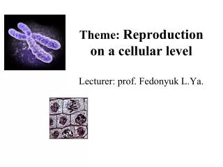
Theme: Reproduction on a cellular level L ecturer : prof. Fedonyuk L.Ya. Plan of lecture:. Structure of nucleus The levels of organization of eukariotic chromosomes. С hromosomes types. Normal human karyotype characteristics. Cell (mitotic) cycle, its stages. Mitosis, its stages.

E N D
Theme: Reproduction on a cellular levelLecturer: prof. Fedonyuk L.Ya.
Plan of lecture: • Structure of nucleus • The levels of organization of eukariotic chromosomes. • Сhromosomes types. • Normal human karyotype characteristics. • Cell (mitotic) cycle, its stages. Mitosis, its stages. • Cytological and cytogenetically characteristics of meiosis.
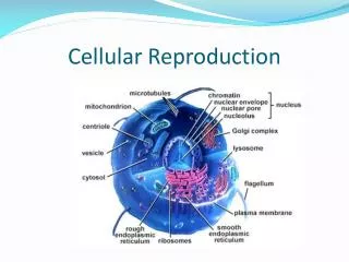
Nucleus consists of • nuclear envelope • nucleolus • nucleoplasm • chromatin (chromosomes)
Nuclear envelope • surrounds the nuclear material • consists of two parallel membranes separated from each other by a narrow perinuclear cisternae • is perforated at intervals by openings called nuclear pores
Nuclear Membrane or Envelope - two membranes which form the nucleus, is porous. Allows RNA to leave nucleus.
Assembly and Disassembly of Nuclear Envelope • Nuclear envelope is a cell cycle dependent structure that disperses at the onset of mitosis (late prophase) and reassembles around the reforming nucleus in the late telophase. • The correlations of breakdown of the nuclear envelope, formation of chromosomes and mitosis are essential for cell division and the ability of cells to divide in an orderly manner.
Nucleoplasm • is the portion of the protoplasm that is surrounded by the nuclear envelope • is consists of a matrix and various types of particles.
Whole Mount Electron Microscopy Demonstrating Fibrogranular Structure of the Internal Nuclear Matrix
Nucleolus • is a well-defined nuclear inclusion (sometime more than one) • is present in the cells that are actively synthesizing proteins • become detectably only when the cell is in interphase • is involved in the synthesis of rRNA and its assembly into precursors of ribosomes
Chromatin • is double-stranded DNA complexed with histones and acidic proteins • is responsible for RNA – synthesis, resides within the nucleus in two forms: heterochromatin and euchromatin
Heterochromatin • is a highly condensed portion of chromatin • is visible in light microscope • appears in the light microscope as basophilic clumps of nucleoprotein • is not transcribed into RNA
Euchromatin • isn’t condensed portion of chromatin during interphase • from which RNA is transcribed, its genes can be activated, is transcriptionally active, mostly encodes proteins • does not visible in light microscope.
Levels of organization of eukaryotic chromosomes: 1. The DNA is associated with basic proteins called histones to form nucleosomes, each of which consists of 8 histones bead with DNA wrapped around it. 2. The nucleosomes are organized into large coiled loops held together by nonhistone scaffolding proteins. 3. The chromonema is a single double-stranded DNA molecule with a protein coat
Chromosome = DNA (deoxyribonucleic acid) + associated proteins (mainly histones) = “packaged” DNA
Organization of eukaryotic chromosomes • The chromosomes have already doubled, and each now consists of two identical sister chromatid • The chromatid is composed of a very fine filament, called as chromonema • The two chromatids remain attached to each other at a point of primary constriction, the centromere. • The centromere is a specific DNA sequence of about 220 nucleotides, to which is bound a disk of protein called a kinetochore. • It is a place, where the spindle fibers attach during cell division. • Regions on either sides of centromere are called arms. • The long arm of a chromosome is designated “q” and the short arm – “p”.
Metaphase chromosome structure 1 - long arm 2 - short arm 3 - centromere 4 - secondary constriction 5 - satellite 6 - chromatids
Types of chromosomes 1 – Metacentric (the centromere divides it into two equal arms) 2 – Submetacentric (the centromere is slightly displace from the center of chromosome) 3, 4 – Acrocentric (the centromere establishes one long arm and one short arm) а – centromere б - secondary constriction
Karyotype • is a diploid number of chromosomes • is represented in humans by the 22 pairs of autosomes and the 1 pair of sex chromosomes (either XX or XY) totaling 46 chromosomes • Pair of chromosomes, with the same gene loci in the same order, are known as the homologous chromosomes. • The chromosomes of each pair have characteristic size and shape. • An ideogram is a karyotype, which displays chromosomes arranged in pairs in descending size order. • 2n=44a+XX(femalekaryotype) • 2n=44a+XY(malekaryotype)
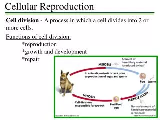
Two important characters of living organisms, Growth and Reproduction are due to Cell division. • Cell division involves two phases • 1. Division of nucleus. • 2. Division of cytoplasm. • Two major types of cell division • 1. Mitosis - similar daughter cells (2n) • 2. Meiosis - Haploid/gametes (n)
The cell cycle • Covers a time from one division of cells till other division or destruction (perish) of cell • It has two major stages: • 1)mitosis • 2) interphase
Cell Cycle: Interphase • Before mitosis • Time of high metabolic activity • DNA replicated and synthesized • Three phases: G1, S, and G2 • G1(gap 1)- longest stage of cell cycle, RNA, protein sysnthesis • S (synthesis)- DNA replicated , 2 chromatids per chromosome, chromatids genetically identical • G2(gap 2)- RNA synthesis, not well understood
Thehuman cell cycle DNA synthesis synthesis S phase G1 phase Rapid growth and preparation for DNA synthesis G2 phase Growth and preparation for cell division M phase Mitosis
Cell Cycle: Mitosis • Process of cell division(nuclear division) which produces daughter cells genetically identical to the parent cell • Four Phases (P-M-A-T): prophase, metaphase, anaphase, and telophase. • Upon completion of the phases of mitosis (nuclear division) the cell “officially” divides into two by a process called cytokinesis - division of cytoplasm
Interphase Not part of mitosis DNA is replicated chromosomes start to condense
Prophase *Chromosomes coil and condense further. *Nucler membrane breaks down/ disappears. *Microtubules increase in number, spindle apparatus forms.
Metaphase *Nuclear membrane completely disappeared *Chromosomes move to equator of cell - begin to line up *Chromosomes attach to spindle via kinetochore
Anaphase *Movement of chromosomes via microtubules to opposite sides of the cell. One chromatid to one end the other Chromatid to the opposite end
In anaphase, the sister chromatids separate. • Two daughter cells • Each has a complete and identical set of chromosomes
Telophase *Genetically identical info at each pole *Spindle fibers disappear *Chromosomes uncoil *Nuclear envelope reforms around
Metaphase, Anaphase, Telophase METAPHASE ANAPHASE Metaphaseplate Nuclearenvelopeforming Spindle Daughterchromosomes
Cytokinesis Cytokinesis - is separate from mitosis, = pinching of cell/divison of cytoplasm. Mitosis + Cytokinesis result in two identical daughter cells.
Mitosis: • Interphase: No morphological changes, Replication of DNA and organelles. • Prophase: Visible chromosomes • Metaphase: equatorial plate formation • Anaphase: Separation of chromosome pairs • Telophase: Two separate nuclei formation. • Cytokinesis: Separation of daughter cells.
Molecular Basis of Carcinogenesis • Genes control cell division by cytokines. • Four classes of regulatory genes. • Promotors – Proto-oncogenes • Inhibitors – Cancer-suppressor genes – p53 • Genes regulating Apoptosis. • DNA repair genes.
Meiosis • Cell division which results in halpoid “sex” cells (i.e., egg and sperm) • One replication of the genetic material (DNA) during interphase, but two nuclear divisions (meiosis I and meiosis II). • Results in haploid (N) cells (= gametes in animals) from an initial diploid (2N) cell • Very similar to mitosis except that the cells produced are not genetically identical.
Meiosis Meiosis I (reduction) Prophase I is divided into the following five stages: • Leptotene -the chromatin condenses into the visible chromosomes, each of which contain two chromatids joined at the centromere • Zygotene - homologous maternal and paternal chromosomes pair and make physical contact (synapsis), forming a tetrad
. Prophase I • Pachytene -the chiasmata are formed and crossing over (random exchanging of genes between segments of homologous chromosomes) occurs – an event that is crucial for increasing generic diversity • Diplotene - the chromosomes continue to condense and chiasmata can be observed, indicating where crossing over has taken place • Diakinesis - the nucleolus disappears, chromosomes are condensed maximally, and the nuclear envelope disappears
Metaphase I • Spindle formation is complete • Bivalents are aligned at equator • Kinetochore microtubules of the homologues point to opposite poles Anaphase I • Homologues separate and move toward opposite poles • Cytokinesis begins