Anatomical Terminology
210 likes | 812 Views
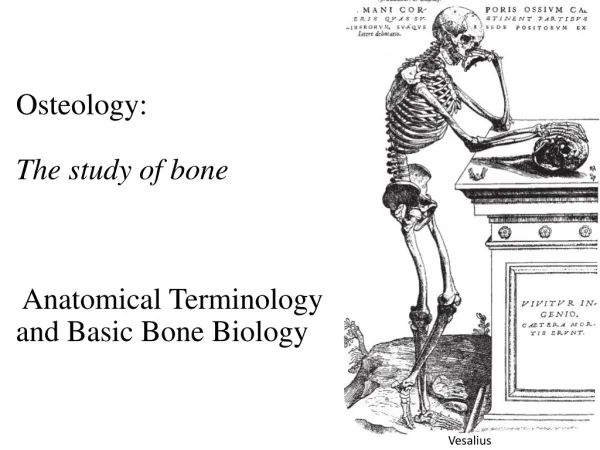
Anatomical Terminology. Lesson 2 Human Anatomy. Body Positions. Anatomical Position The body is standing erect, facing the observer, with the head level and the eyes facing directly forward. The feet are flat on the floor and facing forward.

Anatomical Terminology
E N D
Presentation Transcript
Anatomical Terminology Lesson 2 Human Anatomy
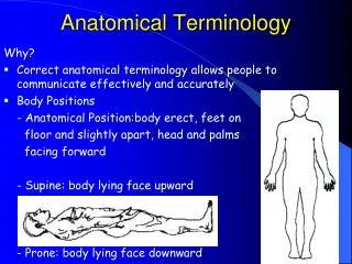
Body Positions • Anatomical Position • The body is standing erect, facing the observer, with the head level and the eyes facing directly forward. • The feet are flat on the floor and facing forward. • The arms are at the sides with the palms turned forward. • Reclining positions: • Prone – the body is lying face down • Supine – the body is lying face up
Regional Names • Head – the skull and face • Neck • Trunk – the chest, abdomen, and pelvis • Upper limbs – shoulder, armpit, arm, forearm, wrist and hand • Lower limbs – buttock, thigh, leg, ankle, and foot
Directional Terms • These terms are used to describe the position of one body part relative to another body part. • Anterior – nearer to or at the front of the body • Posterior – nearer to or at the back of the body • Superior – toward the head or the upper part of a structure • Inferior – away from the hear or the lower part of a structure • Medial – nearer to the midline of the body • Lateral – farther from the midline of the body
Intermediate – between two structures • Ipsilateral – one the same side of the body as another structure • Contralateral – on the opposite side of the body from another structure • Proximal – nearer to the attachment of a limb to the trunk; nearer to the origination of a structure • Distal – farther from the attachment of a limb to the trunk; farther from the origination of a structure • Superficial – toward or on the surface of the body • Deep – away from the surface of the body
Planes and Sections • Planes are imaginary flat surfaces that pass through the body parts. • A section is one flat surface of a three-dimensional structure or a cut along a plane. • Sagittal plane – vertical plane that divides the body or organ into right and left sides. • Midsagittal or median plane – vertical plane that divides the body or organ into equal right and left sides. • Parasagittal plane – vertical plane that divides the body or organ into unequal sides.
Frontal or coronal plane – divides the body or organ into anterior and posterior portions. • Transverse plane – divides the body or organ into superior and inferior portions. Also called a cross-sectional or horizontal plane. • Oblique plane – passes through the body or organ at an angle between the transverse plane and either a sagittal plane or a frontal plane.
Body Cavities • Body cavities are spaces within the body that help protect, separate, and support internal organs. • Cranial cavity: formed by the cranial bones (skull); contains the brain • Vertebral cavity: formed by the vertebral bones; contains the spinal cord • The meninges are three layers of protective tissues that line the cranial and vertebral cavities.
Thoracic cavity: formed by the ribs, muscles of the chest, the sternum, and the thoracic portion of the vertebral column. • Pericardial cavity: fluid-filled space that surrounds the heart. • Pleural cavities: two cavities that surround the lungs. • Mediastinum: central portion of the thoracic cavity that is between the lungs and extends from the sternum to the vertebral column and from the neck to the diaphragm. It contains the heart, esophagus, trachea, thymus, and several large blood vessels.
Diaphragm: a large muscle that separates the thoracic cavity from the abdominopelvic cavity. • Abdominopelvic cavity: extends from the diaphragm to the groin and is enclosed by the abdominal wall and the bones and muscles of the pelvis. • Abdominal cavity: contains the stomach, spleen, liver, gallbladder, small intestine, and most of the large intestine. • Pelvic cavity: contains the urinary bladder, portions of the large intestine, and the internal organs of the reproductive system. • Organs inside the thoracic and abdominopelvic cavities are called viscera.
Membranes of the Thoracic and Abdominal Cavities • Serous membranes • Thin, slippery, double-layers membranes that cover the viscera and also line the walls of the thorax and abdomen. • Parietal layer – lines the cavity walls • Visceral layer – covers and adheres to the viscera • Serous fluid between the two layers reduces friction allowing the organs to slide somewhat during movement.
Types of Serous Membranes • Pleura: serous membrane of the pleural cavities (around the lungs) • Pericardium: serous membrane of the pericardial cavity (around the heart) • Peritonium: serous membrane of the abdominal cavity
Abdominopelvic Regions • The abdominopelvic cavity can be further divided into nine regions by placing two line, tic-tac-toe fashion, across the area. • The top line is placed just underneath the ribs and is called the subcostal line. • The bottom line is placed just inferior to the top of the pelvic bones and is called the transtubercular line. • The left and right midclavicular lines are drawn through the midpoints of the clavicles just medial to the nipples.
The Nine Regions • Top right - Right hypochondriac region • Top middle – Epigastric region • Top left – Left hypochondriac region • Middle right – Right lumbar region • Middle – Umbilical region • Middle left – Left lumbar region • Lower right – Right inguinal (iliac) region • Lower middle – Hypogastic (pubic) region • Lower left – Left inguinal (iliac) region
Abdominal Quadrants • The abdominopelvic region can also be divided into four quadrants by drawing a vertical and horizontal line through the umbilicus (belly button). • Right upper quadrant • Left upper quadrant • Right lower quadrant • Left lower quadrant