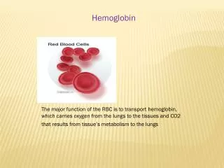
HEMOGLOBIN
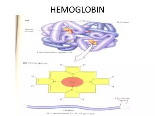
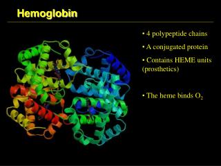
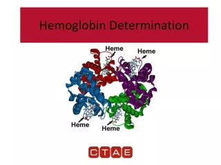
HEMOGLOBIN. Heme is the prosthetic group of hemoglobin, myoglobin, & cytochromes MVMVMPPM. Structure of Heme. Normal Hemoglobin. HEME-CONTAINING PROTEINS Hemoglobin Myoglobin Cytochromes Catalase Some peroxidases. Synthesis of Hemoglobin. GLYCINE + SuccinylCoA.

HEMOGLOBIN
E N D
Presentation Transcript
Heme is the prosthetic group of hemoglobin, myoglobin, & cytochromes MVMVMPPM. Structure of Heme
HEME-CONTAINING PROTEINS Hemoglobin Myoglobin Cytochromes Catalase Some peroxidases
GLYCINE + SuccinylCoA ALA synthase d-aminolevulinic acid(ALA) ALA dehydratase Porphobilinogen(PBG) PBG deaminase hydroxymethylbilane Uroporphyrinogen III cosynthase uroporphyrinogen III Uroporphyrinogen decarboxylase coprophyrinogene III Coproporphyrinogen oxidase Protoporphyrinogene IX Protoporphyrinogen oxidase protoporphyrin IX Ferrochelatase Heme
REGULATION OF ALA SYNTHASE • Down regulated Heme • UP regulated Barbiturates, Steroids (e.g. testosterone) • These drugs are metabolized by the microsomal cytochrome P450 mono-oxygenase system, a heme- containing protein.
LEAD TOXICITY Inhibits multiple enzyme reactions including those involved in heme biosynthesis (PBG synthase & ferrochelatase)Binds to any compound with a sulfhydryl group One symptom of lead toxicity is increases in 5-ALA without concomitant increases in PBG
HAEMOGLOBINOPATHIES • Sickle cell anemia (Hb S) • Hemoglobin C disease (Hb C) • Hemoglobin SC disease (Hb S+ Hb C) • Thalasemia
Sickle cell anemia A 10-years-old African American male presented to ER with complain of pain "all over his body.“ His mother brought him into the ED at 4 pm .She reported that the pain began early that morning and had "gotten worse." She reported that it was not relieved by his usual doses of ibuprofen. He was medicated with strong IM pain killer. He reported minimal pain relief after receiving the medication. He reported that the slight relief was short-lived, and he continued to complain of unbearable pain through the night. His past history is significant with many such hospital admissions and history of repeated chest infections and a non healing ulcer on his right ankle. Family History: History of similar episodes of pain crisis and chest infection in two of the 5 siblings.
Sickle cell anemia Examination: Pale appearing child in agony oriented in time, space and person having a chronic ulcer on right ankle. Cardiovascular System: Moderate tachycardia, grade II/VI systolic murmur heard best over the upper left sternal border. Gastrointestinal Tract: Abdomen: Moderate hepatosplenomegaly. Complete Blood Count: Hb: 5gm/dl, TLC: 12,000/ul, Platelet count: 150,000/ul. Reticulocyte Count: 12%. Peripheral Film: Moderate poikilocytosis, anisocytosis, hypochromia, polychromasia target cells, many fragmented and sickle red cells. Special investigations Sickle Screening Test: Positive HbS: 70%, HbF: 13%, HbA: 17%
Sickle cell anemia (Hb S disease) Homozygous recessive (2 mutant genes that codes forb-chains of globin)-----bS ---------α2b2S (Hb S) Valine replaces glutamate in the 6th position of b-chains • Common in African blacks • Confers resistance against malaria • Hb crystallizes and take sickle shape under hypoxic conditions • Increased RBC Sequestration
THALASSAEMIA • An 8 month old boy was brought by his parents with complaints of lethargy, marked pallor, inactivity and abdominal distension. Eight month old infant presented with the marked pallor and growth failure. There is also history of change in facial appearance. Initially symptoms were less marked. But now they have progressed further. • Family History: History of death of sibling at the age of 15 months diagnosed as deficiency of blood
Examination: Pale appearing, inactive toddler • Mild tachycardia as above, grade II/VI systolic ejection murmur heard best over the upper left sternal border. • Moderate hepatosplenomegaly Complete Blood Count: Hb: 5gm/dl, TLC: 18,000/ul, Platelet count: 150,000/ul. • Reticulocyte Count:10%. • Peripheral Film:Marked poikilocytosis, anisocytosis, microcytosis, hypochromia, polychromasia target cells, many fragmented red cells. • Radiology: X-ray skull show crew cut appearance and maxillary prominence Special investigations HbF: 90%, HbA: 08%, HbA2: 02%
THALASSAEMIA • Beta-thalassemias are a group of hereditary blood disorders characterized by anomalies in the synthesis of the beta chains of hemoglobin resulting in variable phenotypes ranging from severe anemia to clinically asymptomatic individuals. • The total annual incidence of symptomatic individuals is estimated at 1 in 100,000 throughout the world and 1 in 10,000 people in the European Union. 1.5% of the global population (80 to 90 million people) are carriers of beta thalassemia, with about 60,000 symptomatic individuals born annually, the great majority in the developing world.
THALASSAEMIA • Thalassemia Major,"Cooley's Anemia" and "Mediterranean Anemia" • Thalassemia Intermedia and Thalassemia Minor also called"beta-thalassemia carrier", • “Beta-thalassemia trait or"heterozygous beta-thalassemia".
THALASSAEMIA β-Thalassemia • Reduced or absent synthesis of globin chains • 2 copies of β-globin gene on chromosome 11 • β0 No globin chain synthesis • β+ Some globin chain synthesis • β+/β+ Homozygote have anemia of variable severity • β+/β0 CompoundHeterozygote tend to be more severely affected • β0/β0 Homozygote have severe disease
THALASSAEMIA • Excess β-chains form a homotetramer, HbH(Useless for delivering oxygen because of high oxygen affinity) • Inclusion bodies (HbH precipitates trapped and destroyed in the spleen) • Ineffective erythropoiesis: Precipitated α-chains unable to form a stable tetramer
THALASSAEMIA • β -Thalassemia Minor (Make some β-chains. No treatment required) • β -Thalassemia Major (Seemingly healthy at birth , but severely anemic, usually first or second year of life due to ineffective erythropoiesis) • Skeletal changes as a result of extramedullary hematopoiesis • Iron chelation therapy and Bone marrow replacement
THALASSAEMIA α-Thalasemia Deletion mutations 4 copies of the α-Globin gene (2 on each chromosome 16) • Silent carrier of α-Thalasemia: One of the four gene is defective no physical manifestation • α -Thalasemia trait: 3 α-globin genes are defective (Hb β4 disease)—Mild to severe hemolytic anemia • (Hb Bart (γ4 disease)—All 4 α-globin gene defective . Hydrops fetalis