Download
1 / 33
440 likes | 1.21k Views
Bone Density and the DXA Scanner. Pamela Coates. Trabecular & Corticoid. Trabecular. Trabecular more metabolically active Bone loss leads to thinning and perforation of trabecular plates Osteoporotic fractures occur at sites with at least 50% trabecular bone. Trabecular Bone.

E N D
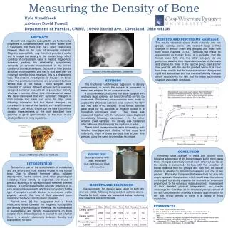
Bone Density and the DXA Scanner Pamela Coates
Trabecular • Trabecular more metabolically active • Bone loss leads to thinning and perforation of trabecular plates • Osteoporotic fractures occur at sites with at least 50% trabecular bone
Trabecular Bone Highlighted areas have at least 50% trabecular bone
Peak Bone Density • Peak bone density is achieved by late 20s/early 30s • Bone loss after skeletal maturity is about 1% per annum in both sexes • Greater loss in woman for around 3 years following menopause
Osteoporosis occurs in: • 1 in 3 postmenopausal women • 1 in 10 men over 50
Classification of Osteoporosis • Primary • Type 1 (postmenopausal) • Type II (senile) • Idiopathic • Secondary • Endocrine (hyperthyroidism, Cushing’s) • Gastrointestinal (coeliac TPN Crohn’s) • Rheumatologocal (RA ankylosingspondylites) • Malignancy • Drugs (corticosteroids heparin)
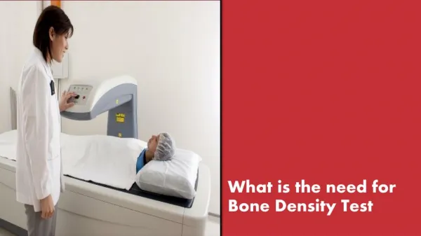
What is DXA • DXA stands for :- Dual Energy X-Ray Absorptiometry • DXA measures, it is NOT an imaging machine • Measurements are taken from the axial skeleton
DXA Scan of Forearm Performed if either of other preferred sites are unusable or in the case of certain conditions Endocrine or gastrointestinal
Total-body scan Shows body composition Used for research Occasionally used to monitor weight – fat loss, muscle gain
Collapsed Vertebra Scan of Lateral spine Scan showing collapsed vertebra Recently approved for the detection of aortic calcification
T-score and Z-score T-score is compared to a population at peak bone density Z-score is compared to an age, sex and ethnicity matched population
T-score T-score- compared to mean of population at peak bone mass T-score >-1 Normal T-score -1 to -2.4 Osteopaenic T-score <-2.5 Osteoporotic
X-rays at two energy peaks are passed through the patientDifferent tissues adsorb differing amounts of each wave lengthDensity is calculated by using simultaneous equation on these amounts How does it work?
There are two manufacturers of DXA Equipment General Electric Company-GE Healthcare Lunar Prodigy Hologic Discovery
Incompatibility The two scanners should not be mixed Patient should continue to be scanned on the original machine They work in different ways
Functionality GE machines K-edge filtration with rare earth filter Hologic machines Alternates between 2 Kvs
Simples! The Math WHERE
Fan Beams GE narrow fan beam Hologic fan beam They have different edge detection GE records lower scores than Hologic
Why do we need a DXA in the breast unit Oestrogen has a beneficial effect on bone density – inhibiting osteoclasts and promoting osteoblasts Without oestrogen this is reversed leading to loss of bone Post menopause oestrogen is converted from androgens by the enzyme aromatase
Aromatase Inhibitors ARIMIDEX (anastrazole) AROMASIN (exemestane) FEMARA (letrozole)
Aromatase Inhibitors As bone density will increase when aromatase inhibitors are prescribed, treatment for loss of bone is given when the T-score is -2 and not the more usual -2.5
Gonadorelin Analogues Gosrelin (zoladex) Busrelin Leuporelin Prostap
Mortality Rate Mortality is increased by 20% in the first year after a hip fracture
UK Annual Costs Annual cost of treating osteoporotic fractures in UK is in excess of £1.73 billion Annual cost of treating coronary heart disease in the UK is £1.75 billion
The lifetime expectancy of a fracture at age 50 is 40% for womenThe likelihood of this will rise for those treated with aromatase inhibitors!”
References Fundamentals of Bone Density (NOS 2002 ATC Study WWW.breastcancer.org WWW.icmri.com www.ch.ac.uk www.osteoporoticdiagnosticcenter.org