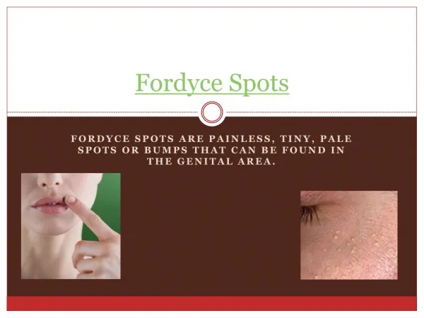
Fordyce Granules
Fordyce Granules. A common condition in which small yellow dots are observed in the oral mucous membrane; these represent misplaced sebaceous glands; once properly diagnosed, no treatment is necessary. Fissured Tongue.

Fordyce Granules
E N D
Presentation Transcript
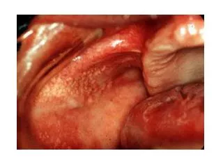
Fordyce Granules • A common condition in which small yellow dots are observed in the oral mucous membrane; these represent misplaced sebaceous glands; once properly diagnosed, no treatment is necessary
Fissured Tongue • A condition of probable developmental etiology in which cracks are observed in the tongue dorsum; food debris and Candida albicans colonies may form in the fissures; once properly diagnosed, no treatment is necessary.
Varix • A fairly-common condition of older people in which distended blood vessels are observed on the lingual tongue surface; once properly diagnosed, no treatment is necessary.
Torus Palatinus • A hamartomatous overgrowth of bone from the midline of the maxilla.
Torus Mandibularis • A hamartomatous overgrowth of bone bilaterally from the lingual surface of the mandible.
Exostosis • Overgrowth of bone (like tori) from a site other than the maxillary midline or bilateral lingual mandible; commonly occur in the buccal maxilla adjacent to the bicuspid teeth.
Nasopalatine duct cyst • A common true jaw cyst appearing as a radiolucency in the maxilla midline just lingual to the central incisor teeth (in the incisive canal); surgical removal will cure this cyst.
Oral Lymphoepithelial Cyst • Uncommon lesion that develops in lymphoid tissue in the oral pharynx including the palatine tonsils, lingual tonsils, and pharyngeal adenoids. They may also arise within accessory lymphoid tissue on the floor of the mouth, ventral surface of the tongue, and soft palate. These small (rarely exceed 1cm) submucosal cysts have a yellow or white appearance and feel firm on palpation. They contain creamy or cheesy keratinous material and are usually asymptomatic except when they are traumatized of become infected. They may occur at any age but are most prevalent in young adults. Cysts rarely recur after surgical excision.
Amelogenesis Imperfecta • The designation "amelogenesis imperfecta" refers to inherited defects in enamel formation. Several forms are recognized based on their pathogenesis and severity. • Clinically, affected enamel may be thinner than normal (generalized hypoplastic form), may be of normal thickness but lacks strength (hypocalcified form), or may be pitted (hypoplastic pitted form).
Periapical Cyst • This cyst is a direct sequela of inflammation of the pulp which has extended into the adjacent periapical tissues. • Treatment consists of endodontic therapy or extraction of the associated tooth with curettage of the cyst.
Acute Necrotizing Ulcerative Gingivitis • Poor oral hygiene combined with serious life stress and possibly nutritional deficiencies. Aka: trench mouth. It can be painful, and it is characterized by areas where the gum tissue has become so inflamed that it has become necrotic. These areas will be small ulcers, and will be grayish in color, and will tend to slough off. The tissue will be generally swollen, and where it isn't dead, it will bleed very easily.
Pseudomembranous Candidiasis • A common fungal infestation of the oral cavity in the immunocompromised or antibiotic-treated patient. Appears as milky white areas on the oral mucosa; lesions wipe-off leaving red (erythematous) base. Responds well to antifungal agents.
Angular Cheilitis • Erythema, fissuring, and scaling at the angles of the mouth. • Often in an older person with reduced vertical dimension of occlusion • Caused by C. albicans, S. aureus (in kids – Rx: OTC triple antibiotic ointment), often both.
Primary Herpetic Stomatitis • Effected mucosa develops numerous pinhead vesicles which collapse to form small, red lesions which enlarge and develop central areas of ulceration covered in yellow fibrin. • Adjacent ulcerations may coalesce to form larger, shallow, irregular-shaped ulcerations. • Distinctive punched-out erosions along the midfacial free gingival margins
Recurrent Herpes Labialis • “cold sore” or “fever blister” • Most common site for recurrence – vermillion boarder and adjacent skin of the lips • Small, erythematous papules; clusters of fluid filled vesicles – rupture and crust within 2 days.
Recurrent Intraoral Herpes Simplex • Multiple coalescing ulcerations on FIXED MUCOSA
Herpes Zoster • Recurrent infection, often after several decades • Virus was latent in the dorsal spinal ganglia • Recurrent intraoral: • Unilateral, severe pain • Looks like recurrent herpes simplex
Linea Alba • Normal variation in the buccal mucosa that appears as a white line beginning at the corners of the oral cavity and extending posteriorly at the level of the occlusal plane. It is composed of keratinized oral mucosa.
Cheek Chewing • Hyperkeratosis caused by habitual chewing on the cheek
Amalgam Tattoo • A bluish-black or gray macular lesion of the oral mucous membrane caused by accidental implantation of silver amalgam into the tissue during tooth restoration or extraction.
Antral Pseudocyst • The antral pseudocyst is a common and well-documented finding on a panoramic radiograph. Most are discovered during routine radiographic examination and have little clinical significance. The antral pseudocyst should not be confused with the mucocele of the sinus. The antral pseudocyst is believed to be caused by an inflammatory exudate that accumulates under the maxillary sinus mucosa and results in a sessile elevation.
Aphthous Lesion • A very common self-limiting, episodic disease of unknown origin manifesting as one or several painful ulcers on the lining mucosa; the ulcers heal in 7-10 days with or without treatment.
Major Aphthous Stomatitis • Major aphthae are more serious ulcerative disease of unknown etiology manifesting as large, deep, painful ulcers that leave scars; they recur more often and persist longer than minor aphthae; treatment consists of promoting healing and alleviating pain.
Cinnamon Stomatitis • Contact stomatitis from artificial cinnamon flavoring (concentrations of the artificial flavoring are up to 100 times that in the natural spice). Most common in chewing gum, candy, toothpaste.
Dentifrice Stomatitis • Superficial epithelial sloughing, no pain or bleeding, a reaction to detergent in toothpaste