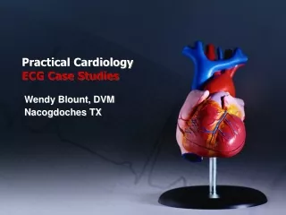
ECG Case Studies
ECG Case Studies. Moosa Kalla. Case 1. 52 yr old man No Hx of IHD Known HPT on Rx Presents with acute onset chest Initial ECG normal Cardiac enzymes normal Admitted for observations. ECG 24 Hrs post admission. ECG findings. Rate: 50 Rythym: sinus PRI: normal QRS: <0.12

ECG Case Studies
E N D
Presentation Transcript
ECG Case Studies Moosa Kalla
Case 1 52 yr old man No Hx of IHD Known HPT on Rx Presents with acute onset chest Initial ECG normal Cardiac enzymes normal Admitted for observations
ECG findings • Rate: 50 • Rythym: sinus • PRI: normal • QRS: <0.12 • : Rwave progression normal • ST seg: biphasic Twaves V2-V5 • slight STE V1 • No Q waves • AVR normal
Management Diagnosed with Wellen’s Syndrome Coronary angiogram showed 95% stenosis of LAD Percutaneous angioplasty and stinting performed Patient discharged 3 days later
Wellen’s Syndrome 1982 Wellen’s et al first published ECG criteria for subgroup of pt. with AMI Later came to be known as Wellen’s syndrome Wellen’s syndrome is a pre-infarction stage of coronary artery disease Recognition of this ECG pattern allows identification of pt with severe LAD disease and hence at risk of anterior wall MI
Charecteristics of Wellen’s Sx Charecterised by Bi-phasic or T wave inversion in precordial leads Typically caused by critical stenosis in proximal LAD The charecteristic ECG pattern often develops while pt is pain free During chest pain ST-segemnet-T-wave abnormalities normalize or develop into ST-segment elevation
Case 2 28 year old man c/o lightheadedness and shortness of breath,than collapses On scene is PEA, CPR instituted and intubated Arrives in ED 15min post collapse ECG showed fine VF Defib at 200J and ECG redone at 2min
ECG FINDINGS Rate: 75 Rhythm: sinus PRI: normal Axis: normal QRS:RSR V1 V2, Incomplete RBBB ST elevation V1 V2, downsloping
Brugada syndrome • Described by Brugada and Pedro 1992 • Frequent cause of death in pt. with normal hearts • Also a cause of sudden death in athletic population • More frequently diagnosed in males of South East Asian descent • Charecterised by ECG abnormalities in V1 to V3: i ) incomplete RBBB • ii) ST segment elevation
) Caused by a reduction of sodium current across cardiac sodium channels ST elevation thought to be due to rebalancing of currents active at end of phase 1 Definitive treatment is by placement of Internal Cardio-defibrilator(ICD ) Mortality at 10yrs is 0%for ICD and 26% for pharmocological agents(amiodorone,B-blockers Mortality at 10yrs is 0%for ICD and 26% for pharmocological agents(amiodorone,B-blockers
Case 3 40yr old man, 2d HX intermittent chest pain Hx of smoking, hyperlipidaemia and PUD O/E T 37.5 BP 140/80 P100 Heart sounds distant ,no cardiac or pleural rubs ECHO and CXR normal
ECG Findings Rate:140 Rythym: sinus PRI: normal PR seg: elevation aVR, : depression ii V5 V6 Axis: normal QRS: <.012 ST seg: concave STE I II III V4-V6 No reciprical changes
LAB findings Trop t negative WCC 12.5 ESR 50 Urgent angiography showed healthy coronary arteries
Pericarditis Pericarditis syndrome caused by inflamation of pericardium There is increased vascular permeability, vasodilation and transudation Patient presents with sharp central chest pain worse with inspiration and recumbency Pain may radiate
. O/E pericardial friction rub is a pathognomic finding,best heard in expiration,heard 50% of times Distinct ECG findings: i) Concave ST elevation ii) PR seg depression iii) widespread STE not corresponding to any arterial territory iv) Absence of reciprocal changes and Q waves v) Possible presecnce of low voltages (STE II>STE III strongly favours acute pericarditis;STE III>STE II strongly favours AMI
Case 4 58 yr old man, 45min severe chest pain Grey sweaty,nauseous,SOB,anxious Clinically RR 16 BP 135/75 P 75 Heart sounds normal, no mumurs
ECG Rate: 80 Rythym: sinus PR: normal QRS: LBBB ST seg: global discordance : concordance V4 1 mm
Sgarbossa criteria LBB on ECG may mask changes of AMI Can delay reognition of AMI and thrombolysis Sgarbossa et al tested criteria for AMI in presence of LBBB Data used from patients enrolled on GUSTO-1 trial These patients had AMI confirmed by enzyme studies
Findings ST segment deviations only ECG findings useful in diagnosisng acute myocardal infarction in the presence of LBBB
Criteria selected The ST changes that were significant are:1.ST elevation > or = 1mm and concordant with QRS.2.ST depression > or = 1mm in v1,v2 or v3.3.ST elevation > or = 5mm and discordant with QRS.
Concept of Con/discordance • Refers to whether the last portion of the QRS complex goes in the same or opposite direction to the T wave • Discordance=opposite=good= secondary • Concordance= same=bad=primary
ECG 5 • Elderly lady,far-east origin • New onset chest pain • Nausea and diaphoresis • Recent severe social stressors
Hospital course • Emergency cardiac catherisatrion… no obstructive coronary artery disease • Patient had haemodynamic profile of cardiogenic shock: • intra-aortic balloon pump • started on vasopressor support
ECHO findings at 24 hours • Moderate to severe systolic dysfunction of LV which is segmental • Only proximal segment of IV septum and anterolateral wall contracting normally • Ballooning of distal ventricle • EF estimated at 20% • Consistent findings of Taka-Tsubo syndrome • Moderate mitral regurgitation
ECG Findings • Rate: 100 • Rythym: sinus • PRI: normal • Axis: left • QRS: narrow • ST seg: STE V-V5 • : biphasic V3-V5 • : inverted V6
Tokatsubo Cardiomyopathy Acute stress cardiomyopathy,described as form of Reversible Left Ventricular Systolic Dysfunction in the absence of coronary artery disease First described in Japan Now global distribution Also known as Broken Heart Syndrome (BHS) Pathogenisis not well understood More common in woman aged 62-75
Presentation Typically triggered by emotional, physical or medical stressors Commonly present with SOB Shock ECG changes of ischaemia
Postulated mechanisms i) cathecholamine-induced induced vent dysfunction(due to stress hormone release) ii)multivessel coronary spasm iii) dynamic left vent outflow tract obstruction
Distinguishing from ACS Features distinguishing SC from LAD territory infarction are: i) Abnormal ST elevation/depression, t wave inversion, raerely Q waves ii) cardiac biomarkers mildly elevated iii) wall motion abnormal on ECHO-large area for single artery involvement iv)Lack of delayed hyperenhancement on MRI with gadolinium
Clinical course Recovery of baseline Left ventricular function within 1-4 weeks Low mortality ranging from 0-8% Diagnosis is mainly by exclusion of ACS NB suspicion of stress cardiomyopathy not sufficient reason to withold treatment for acute ACS…stress cardiomyopathy diagnosed by presence of all 4 criterai listed above
ECG findings • Rate: 66 • Rythym: ventricular paced • Axis: left • QRS: LBBB • :Q waves V1-V6 • ST seg: discordant all leads except V2
ECG • Rate: 66 • Rythym: sinus • Axis: normal • PRI normal • QRS: LBBB • ST seg: STE II III aVF • : reciprocal changes aVL and • V2
Management Aspirin 300mg TNT 2 tabs S Morphine 2.5mg IVI GTN infusion commenced Pain decreased from 8/10 to 6/10 Spontaneously reverted to native rythym