Download
1 / 12
120 likes | 785 Views
Penetrating Keratoplasty vs. Deep Lamellar Keratoplasty in Macular Dystrophy: Case Report. Amit Patel MRCOphth, Harish Nayak MRCOphth, Vinod Kumar FRCSEd(Ophth) Princess of Wales Hospital, Bridgend, UK. The authors have no financial interests with regards to this poster. Introduction.

E N D
Penetrating Keratoplasty vs. Deep Lamellar Keratoplasty in Macular Dystrophy: Case Report Amit Patel MRCOphth, Harish Nayak MRCOphth, Vinod Kumar FRCSEd(Ophth) Princess of Wales Hospital, Bridgend, UK The authors have no financial interests with regards to this poster
Introduction • Penetrating keratoplasty (PK) is commonly performed for macular corneal dystrophy • Traditional teaching guides against lamellar keratoplasty for macular dystrophy due to: • deep stroma/descemet’s membrane involvement • concern about endothelial health • This case describes a patient with macular dystrophy who underwent a PK in one eye and deep lamellar keratoplasty (DLK) in the fellow eye
Case • A 54-year-old man presented with increasing glare and reduced vision affecting both eyes • Best corrected visual acuities (BCVA) were 6/12 OU • Bilateral multiple grey-white stromal opacities consistent with macular dystrophy were noted • Keratoplasty was offered as driving was essential for his occupation
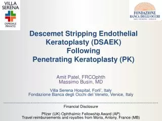
Case - OS • Manual deep lamellar dissection (Melles’ technique) in the left eye revealed a residual hazy bed due to significant descemet’s membrane involvement • The operation was thus converted to a PK (see Fig.1) • Continuous suture was adjusted four months post-operatively
Case - OD • The ‘Big-Bubble’ technique was used to perform lamellar dissection in the right eye • The residual bed was noted to be relatively clear and the DLK completed (see Fig. 2) • Continuous suture was adjusted two months post-operatively
Initial interface haze in the right (DLK) eye had cleared by 3 months. Off axis descemet’s membrane creases were not visually significant. Note the discrete residual opacities (arrow) BCVA at last review (18 months post-op) was 6/6 with spectacle prescription: +0.50/-1.50 x 95 Outcomes Fig 1
The left graft (PK) remained clear. Note the stromal opacities in the residual host rim (arrow) BCVA at last review (24 months post-op) was 6/9+ with spectacle prescription: +3.50/-3.00 x 3 …outcomes Fig 2
…outcomes • Endothelial cell counts were comparable with no significant difference at 14 (OD, DLK) and 26 (OS, PK) months post-op • Subjective & Objective visual acuities were better in the DLK eye OD OS
Discussion • The DLK learning curve and potential newer complications (double anterior chamber, descemet’s rupture) are outweighed by the risks of ‘open sky’ surgery • PK has been shown to offer faster visual recovery than DLK, although no difference in final visual- & contrast acuities has been found
…discussion • Numerous advantages of DLK over PK exist: • Lower rejection rates • 20% rejection rate with 3.5% failure rate has been reported in 229 cases of macular dystrophy undergoing a PK • Ease of re-grafting • Up to 25% recurrence of macular dystrophy in patients with PK has been reported over 7-22 years • Lower endothelial cell loss • Study comparing DLK & PK for various corneal opacities showed lower endothelial cell loss and intraocular pressure rise in the DLK group
Conclusion • DLK may be a superior choice in the surgical management of macular dystrophy and should be considered when the endothelium is healthy
References • Anwar M, Teichmann KD. Big-bubble technique to bare Descemet's membrane in anterior lamellar keratoplasty. J Cataract Refract Surg. 2002 Mar;28(3):398-403. • Melles GR, Rietveld FJ, Beekhuis WH, Binder PS. A technique to visualize corneal incision and lamellar dissection depth during surgery. Cornea. 1999 Jan;18(1):80-6. • Shimazaki J, Shimmura S, Ishioka M, Tsubota K Randomized clinical trial of deep lamellar keratoplasty vs penetrating keratoplasty. Am J Ophthalmol. 2002 Aug;134(2):159-65. • Kawashima M, Kawakita T, Den S, Shimmura S, Tsubota K, Shimazaki J. Comparison of deep lamellar keratoplasty and penetrating keratoplasty for lattice and macular corneal dystrophies. Am J Ophthalmol. 2006 Aug;142(2):304-9. • Vajpayee RB, Tyagi J, Sharma N, Kumar N, Jhanji V, Titiyal JS. Deep anterior lamellar keratoplasty by big-bubble technique for treatment corneal stromal opacities. Am J Ophthalmol. 2007 Jun;143(6):954-957. • Lyons CJ, McCartney AC, Kirkness CM, Ficker LA, Steele AD, Rice NS. Granular corneal dystrophy. Visual results and pattern of recurrence after lamellar or penetrating keratoplasty. Ophthalmology. 1994 Nov;101(11):1812-7. • Al-Swailem SA, Al-Rajhi AA, Wagoner MD. Penetrating keratoplasty for macular corneal dystrophy. Ophthalmology. 2005 Feb;112(2):220-4. • Marcon AS, Cohen EJ, Rapuano CJ, Laibson PR. Recurrence of corneal stromal dystrophies after penetrating keratoplasty. Cornea. 2003 Jan;22(1):19-21. • Akova YA, Kirkness CM, McCartney AC, Ficker LA, Rice NS, Steele AD. Recurrent macular corneal dystrophy following penetrating keratoplasty. Eye. 1990;4 ( Pt 5):698-705.