Download
1 / 17
220 likes | 912 Views
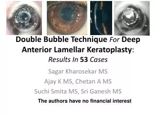
Corneal Densitometry Measurement After Deep Anterior Lamellar Keratoplasty by Oculus Pentacam . Eric Dai MD, Pawan Prasher MD, James McCulley MD, R. Wayne Bowman MD. The University of Texas Southwestern Medical Center, Dallas, TX. Commercial Support Disclosure.

E N D
Corneal Densitometry Measurement After Deep Anterior Lamellar Keratoplasty by Oculus Pentacam Eric Dai MD, Pawan Prasher MD, James McCulley MD, R. Wayne Bowman MD. The University of Texas Southwestern Medical Center, Dallas, TX.
Commercial Support Disclosure • The authors do not have any financial arrangements or affiliations regarding the products and technology used in this study.
Purpose • To correlate changes in corneal densitometry values from Oculus Pentacam with clinical slit lamp examination after deep anterior lamellar keratoplasty (DALK).
Methods • Pentacam images and slit lamp photographs were performed in nine eyes of nine patients at various intervals during the postoperative course after DALK. • Nine eyes of nine patients with no prior corneal surgery served as controls. • The values for peak anterior stromal density and posterior stromal density at the level of the graft interface along the 180 degree axis were chosen for analysis. • These values were subsequently correlated with clinical grading of interface haze using previously published guidelines1.
Clinical Grading of Interface Haze1 • Grade 0 for a completely clear cornea • Grade 0.5 for trace haze seen with careful oblique illumination with slit-lamp biomicroscopy • Grade 1 for more prominent haze not interfering with visibility of fine iris details • Grade 2 for mild obscuration of iris details • Grade 3 for moderate obscuration of the iris and lens • Grade 4 for completely opacification of the stroma
Results • On slit lamp examination, interface haze varied from grade 0.5 to 3 in the study group. • Both anterior and posterior stromal densitometry values in the DALK patients were significantly greater than in the control group (p<0.01).
Conclusion • Corneal densitometry values obtained by the Oculus Pentacam were signifcantly higher in patients with interface haze after DALK. • Corneal densitometry appears to correlate with subjective assessment of corneal interface haze by clinical slit lamp examination in patients undergoing DALK. • The Pentacam may be a useful tool to quantify interface haze in the post operative course of DALK. • Additional study in a larger patient population is needed to further evaluate the utility of the Pentacam in grading interface haze.
References • Fantes FE, et. al. Wound healing after excimer laser keratomileusis (photorefractive keratectomy) in monkeys. Arch Ophthalmol. 1990 May;108(5):665-75.