Download
1 / 33
330 likes | 443 Views
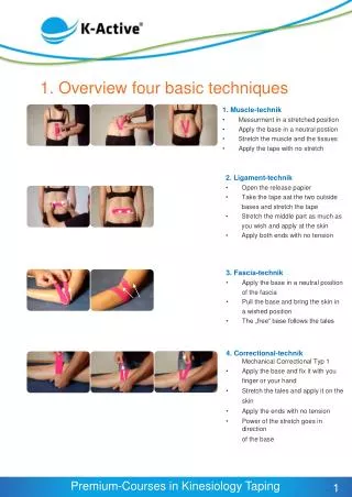
1. Overview four basic techniques. 1. Muscle-technik Messurment in a stretched position Apply the base in a neutral postion Stretch the muscle and the tissues Apply the tape with no stretch . 2. Ligament-technik Open the release papier Take the tape aat the two outside

E N D
1. Overview four basic techniques • 1. Muscle-technik • Messurment in a stretched position • Apply the base in a neutral postion • Stretch the muscle and the tissues • Apply the tape with no stretch • 2. Ligament-technik • Open the release papier • Take the tape aat the two outside • bases and stretch the tape • Stretch the middle part as much as • you wish and apply at the skin • Apply both ends with no tension • 3. Fascia-technik • Apply the base in a neutral position • of the fascia • Pull the base and bring the skin in • a wished position • The „free“ base follows the tales • 4. Correctional-technikMechanical Correctional Typ 1 • Apply the base and fix it with you • finger or your hand • Stretch the tales and apply it on the • skin • Apply the ends with no tension • Power of the stretch goes in direction • of the base 1
1.1. Muscle-technique „Sacrospinale system“ 1 2 3 Picture 1: The lenght of the tape you measure in the stretched position of the are. Picture 2: Apply the base in neutral postition in the area of the sacrum. Picture 3: Stretch the skin and the soft tissues as much as possible (accept pain). 5 6 4 Picture 4: The tails and the ends apply with no stretch in the area of the muscle. Pictures 5/6: Back in neutral position you have to see the Convolutions. • Effects: • Tostrength a weakor a not correctreacticvemuscle • Plus theeffectsoftheconvulutions • The power ofthestrips will go soft back tothebase. 2
1.2. Ligament- or Space-technique „Lower back“ 2 3 1 4 5 • Effects: • Bystretchingthetape, energy will storage in thetape. After theapplicationthisenergy will bring thetapeandthetissues a little in directionofthemiddle. • Depending on thestretchyou will getstabilationorspace. 3
1.3. Ligament- or Paincross 1 2 3 Picture 1: Ligament-technique applyvertical Picture 3: Ligament-Technique apply horizontal Picture 2: The next two techniques do cross in the area of the pain 1.4. Triggerpoint Apply 3-4 small Ligament-techniques Crossed in the area of the trigger point. 4
1.5. Fascia-technique 1 2 Picture 1: Apply the base with no stretch. Picture 2: Pull soft with soft vibrations on the tales to change slightly the base. Apply always during the pulling small parts of the tails.. 3 4 Effects: By pulling the tales the base will following this direction and the skin and the fascia could changed in a „wished“ postion. With this application you will get change of the positioning and tension of the skin, fascia and other tissues. The Y-shape you especially can use for pain points. You can put it in the middles of the Y. 5
1.6. Correctional-technique 3 2 1 Picture 1: Apply the base with no stretch. Picture 2: Fix the base. Now you can stretch the tails from moderate till maximum. Picture 3: Apply the ends with no stretch. Picture 4: Finishes application. 4 Effects: By fixing the base, the power of the tales will come back to the base.(rubber effect). With this application you can change positioning and tension of skin, fascia, soft tissures, but also bones like the patella. With this application you can get more structural and sensitive input for as with the fascia-technique. 6
Screening of the dorsal myofascialchain Generell testsforthe dorsal myofascialsystem/ chain: 1. UpperSpine Test 2. LowerSpine Test 3. SLR orLaseque Test 7
Screening of the ventral myofascialchain Generell testsforthe dorsal myofascial system/ chain: 1. Cervical Extension Test 2. Thorax Test 3. Abdominal Pressure Test 4. Globaltest: Hyperextension of thebody 8
Screening of the diagonal myofascialchain Generell testsforthe dorsal myofaszcale system/ chain: 1. UpperExtremity Test 2. Thorax Test 3. Abdominal Pressure Test 4. Patrick Test 9
2. Lower Spine Test Possible muscles to be tested: M. sacrospinalis M. quadratus lumborum M. iliocostalis lumborum M. psoas major M. illiacus • Patient lies supine and performs an assistive flexion of the entire spine. • The therapist should assess the following especially in the lower back: • Pain at the starting point, during and/or at the end of the movement • 2. Can the patient perform the movement themselves - only partially or full • range of motion possible? • 3. Are there sensitive zones on the skin (soft tissue zones, headache zones, etc) • 4. Flexibility of the skin on the dorsal side • 5. Folding/wrinkles of the tissue on the ventral side • 6. Fascial mobility (in relaxed position) • 7. Check the temperature of the tissue and the energetic radiation • This test is positive, if one or more factors are abnormal. Test in thestandingposition 10
2.1. Sacrospinal muscle system 1 2 3 Picture 1: The lenght of the tape you measure in the stretched position of the are. Picture 2: Apply the base in neutral postition in the area of the sacrum. Picture 3: Stretch the skin and the soft tissues as much as possible (accept pain). 4 5 6 Picture 4: The tails and the ends apply with no stretch in the area of the muscle. Pictures 5/6: Back in neutral position you have to see the convolutions. • Effects: • Tostrength a weakor a not correctreacticvemuscle • Plus theeffectsoftheconvulutions • The power/directionofthestrips will go soft back tothebase 11
Remodelingtechniques Expansions Zones, scars, operation, burningwounds, ... 1 3 2 Pic. 1: Expansion belowthebellybottom Pic. 2: Stretch the Tape gentle ( 25 %) and thenapplyitwith„pressure“ in thetissue. Pic. 3: Completeappication: nowthetissueis a littlemoreinside Shrinkings Zones, scars, operation, burningwounds, ... 4 5 6 Pic. 4: Tissueismoreinside Pic. 5:Stretch thetissueasmuchaspossiblee.gextension, liftthearmsbreath in … and thenthetape will beappliedwithnotension Pic. 6:Completeapplication: nowthetissueis a littlemore outside . Effects: Balancing of bodyshapestogetbetterbodyfunctions 12
3. Upper Spine Test Possible muscles to be tested: M. trapezius M. latissimus dorsi M. levator scapulae M. erector trunci • Patient lies supine and through an assistivemovementgoesinto spinal flexion up to TH12. • The therapistassessesthefollowing: • Pain at the starting point, during and/or at the end of the movement • 2. Can the patient perform the movement themselves - only partially or • full range of motion possible? • 3. Are there sensitive zones on the skin (soft tissue zones, Head • zones etc) • 4. Flexibility of the skin on the dorsal side • 5. Folding/ wrinkles of the tissue on the ventral side • 6. Fascial mobility (in stretched and relaxed position) • 7. Check the temperature of the tissue and the energetic radiation • This test is positive, if one or more factors are abnormal. Test in thestandingposition 13
3.1. M. trapezius (pars descendens) Picture 1: Possible version: Base is applied in neutral over the inferior tip of the acromium. 1 Picture 2: Bring the muscle into maximum stretch: Originally application: lateral flexion and rotation to the opposite side, cervical flexion. Anatomically: Lateral flexion to the opposite side and rotation to the same sid with cervical flexion. 2 Picture 3: Apply the tape along the body of the muscle ending at the base of the hair line. 3 14
4. Thorax Test • Possible Muscle to be tested: • M. rhomboideus major • M. rhomboideus minor • M. sternocleidomastoideus • M. pectoralis minor • M. diaphragma • Mm. intercostales The patient lies supine, the therapist assesses the thorax. Observation: asymmetries, scars, zones, problems with the fascia. Palpation: flexibility of fascia and complications (zones) in this region Pressure test of the ribs and underlying tissue and organs. The test is positive if you find pain, changes to the fascia or skin or other abnormalities. 15
4.1. M. rhomboideusmajor 2 1 Picture 1: Centre of tape is applied medial of the medial border of the scapula (X-shape). Picture 2: Roll both shoulders forward / down and then flex the neck to fully stretch the tissues. Apply the superior strip towards TH2 and the inferior strip to TH5. 3 4 Pictures 4 /5: Apply the lateral strips of tape in the same manner and angle as above. Picture 5: Final application. 5 16
5. Upper Extremity Test • Possible muscles to be tested: • Chest – M. pectoralis major • Shoulder and arm muscles • Muscles of the hand due to large • representation area in our cortex! The patient is seated on a chair. The therapists grips into the palm of the patients hand and passively/assistiv brings the arm into outward rotation (behind vertical line), horizontal abduction. The norm is 0° horizontal abduction and ability to bring shoulder/arm by rotation behind the vertical. The test is positive, if range of movement is not 100%, there is pain or other restrictive factors are noted. Additionally therapist should assess the following: presence and flexibility of scars and fascia anteriorly and posteriorly. Folding of the skin over posterior thoracic area as well as into the posterior arm. 17
5.1. M. deltoideus 1 2 Picture 1: Base: applyin neutral position belowthedeltoidtuberosity. Picture 2: Bring ventral section of deltoid muscle into full stretch and apply tape on anterior border of muscle. Picture 3: Bring dorsal section of themuscleintofullstretch and applythetape on posteriorborder of muscle. 4 3 Picture 4: Final Application. 18
5.2. M. supraspinatus Picture 1: Base isapplied in neutral postion in thearea of the insertion. Will bethere a painpoint, thenthetape will applied in a littledistant of thepoint. 1 Stretch themuscle and thefascia. Applytapealong fossasupraspinatussurrounding of thebodyof themuscle. 5.3. Impingement-Syndrom Picture 3: Fascia-technique. * Base: anterior of shoulder Withjiggling of tape, pull fasciapostriorly and apply (onepossibility). The painpointis in themiddle of the Y 2 • Picture 3: FinishedAppilcation: • M. deltoideusMuscletechnique • M. supraspinatusMuscletchnique • Fasciatechnique 3 19
6. Cervical Extension Test • Possible muscle to be tested: • Mm. scalenii • M. splenius capitis • M. splenius cervicis • M. sternocleidomastoideus • The patient is seated and actively extends the cervical spine. • The therapist assesses the following: • 1. Pain at the starting point, during and/or at the end • of the movement • 2. Can the patient perform movement themselves – only partially or full range of motion • possible? • 3. Are there sensitive zones on the skin (soft tissue zones, Head zones, etc) • 4. Folding of the skin on the dorsal side • 5. Flexibility of the tissue on the ventral side • 6. Fascial movement (in relaxed position) • 7. Check the temperature of the tissue and the • energetic radiation • This test is positive, if one or more factors are abnormal. 20
6.1. Whipplash Picture 1: Base over scapula. Application of tape with 10-15% stretch. Base is fixed and cervical spine in flexion or the positon of pain free. 1 Picture 2: Finished application of first fan 2 Pictrue 3: Finished application 3 21
6.2. Cervialspineandheadache 2 1 • Picture 1: • Muscle-technique • base depending on the testing • e.g. base under the hairline. • Picture 2: • Ligament technique over C7 • depending on the testing. 22
6.3. Epicondylitisrad./uln. hum. Picture 1 - 2: Fasciatechnique (assesdirection!) 1 2 Picture 2 - 3: Muscletechnique (assesdirection!) 3 4 Picture 5: Possiblecombination Cave: Normallythepainpoint will not coverbytape. * 5 23
7. SLR / Laségue Test • Possibel muscles to be tested: • all muscles of the leg and the feed • additional the muscles of the lumbar rigion • at the end point of the movement, also the • other parts of the myofascial backline The patient lays supine with legs in neutral. The therapist performs a SLR gripping from the heel. Observation: pain? when? at what point does the tension in the muscles change, how is the tension in the fascia, especially dorsally (including the lower back)? Are the ventral fascia able to relax / fold during hip flexion? The test is positive, if there is pain, abnormal movement or no normal folding ventrally. 24
7.1. Lymphatic fan - Knee Lymphatic applications always apply accordant to the problems of the patient • Picture 1: Lateral Lymphatic fan • Base proximal to the area of the problem • Apply the tape with different angels of the • knee flex with around 10 % stretch of the tape • Picture 2: Medial Lymphatic fan • Base proximal to the area of the problem • Apply the tape with different angels of • the knee flex with around 10 % stretch of the • tape Additional there could be apply a tape for the scars and the muscle 25
7.2. M. quadriceps (generalapplication) Picture 1: The leg lies extended on the table. Base: below the SIAI in neutral hip / knee position (Full version). Place hip into extention of the side of the bed with knee in flexion. Apply the tape over the first 1/3 of the muscle. 1 Picture 2: Bring leg into hip and knee flexion standing leg on the bed. Apply the tape on the ramaining muscle body allowing the strips of tape to tail off around the patella. 2 Picture 3: Final application. 3 26
7.3. Indicationapplication: Knee 1 2 Picture 1: Muscle technique M. quadriceps. Picture 2: Mech. Correction type 1. Base over Tub. Tibiae, fix base, Knee in max. Flx. Apply tape with 100% stretch towards apex patella. Lay on ends without stretch in direction of the Mm. vastus medialis and lateralis. Picture 3: Final application. 3 4 5 Picture 4 /5: Muscle technique hamstrings out of the standing or lying position. 27
7.4. ApplicationAchilloTendon 1 2 Picture 1: Tendon-Technique Foot in Dorsal extension: Base over the calcaneus Distal tape is applied with sub-maximum stretch over the base of the foot towards the toes. Lay the end on with no stretch. Proximal tape is applied with max. stretch over the length of the achilles tendon (first 1/3rd), 50 % stretch over muscle-tendon section (scond 1/3rd) and no stretch over soleus muscle (last 1/3rd). Picture 2: Muscle-technique Base over calcaneus or in the area of the lower calft; Foot in dorsal extension; 2 straps surround the calf muscles. 4 3 Picture 3: „Stirrup“ Ankle at 90°;Base over calcaneus with no stretch; 2 straps pulled proximal over the ankle joint to support plantar flexion – apply tape behind the joint line. Picture 4: Complete Achilles Tendon tape 1. Base on Calcaneus 2 “Tendon-technique“ for achilles tendon and plantar fascia 2. Muscle-technique for calf muscles 3. Stirrup over plantar ankle joint 28
8. Patrick Test • Possible muscles to be tested: • M. glutaeus maximus/ medius / minimus • Mm. adductores • M. iliopsoas • M. tensor fascia latae • M. sartorius • M. piriformis This test is used when the patient has problems around the pelvis and / or hip. The patient lies supine, the knee on the side to be tested is flexion, the hip in external rotation and is placed on the opposite knee. The therapists performs a passive movement into the end range of motion (OR). The test is positive, if the range is limited by pain, contraction of fascia, scars or muscles. (Always compare both sides!) 29
8.1. M. glutaeusmaximus Picture 1: Base: over greater trochanter For the proximal tape: hip in full extension. Tape is applied along the crista iliaca towards the SIPS. 1 Picture 2: For the distal tape: Hip in full flex. Tape applied around muscle belly towards the apex of the sacrum. 2 Picture 3: Final application. 3 30
9. Abdominal Pressure Test • Possible muscles to be tested: • M. rectus abdominis • M. obliquus externus abdominis • M. obliquus internus abdominis • M. transversus abdominis • M. diaphragma The patient lies supine, the therapist examines the abdominal region. Observation: asymmetries, scars, zones, problems with the fascia. Palpation: fascial mobility and complications (zones) in this region. Perform pressure test of the tissue and the organs – first superficially then with deeper pressure. The abdomen can be divided into 9 zones. This test is positive, if you find pain or changes in the fascia or skin. 31
9.1. M.obliquusexternus 1 2 Picture 1: Knee in Flexion, hip in 45° Flexion, adduction and internal rotation. The lower back is straight, arms elevated. Base: Superior of os pubis, just above hairline. Picture 2: During application patent breaths in. Tape is applied towards the 10th rib. 9.2. M. obliquus internus 4 3 Picture 3: Both knees in Flexion. Rotate both knees to the side of application. Arms elevated. Keep lower back in lordosis. Base: Medial of SIAS. Picture 4: Final application. Tape is applied towards the Proc. Xiphoid. 32
9.3. Sacro-iliacjoint(SI-Joint) Picture 1: 2 tapes are applied, which cross over on effected SI-joint. Application of base depends on what SI-Joint position needs correction - anterior or posterior tiltdifferent applications are possible. 1 9.4. Scars and fibrosis 1 2 Picture 1: Using two base. Picture 2: without a base. Applying crossed pattern over the scars and fibrosis – normally with soft stretch. 9.5. Rib fracture 1 2 Picture 1: Ligament technique over the fractured rib. Ligament technique anterior and posterior of fractured area. Picture 2: Variation: Webcut over the fractured rib. Ligament techniques anterior and posterior of the fractured area. Alternative: Cross two lympathic fans, base under the fracture position 33