Download
1 / 22
220 likes | 411 Views
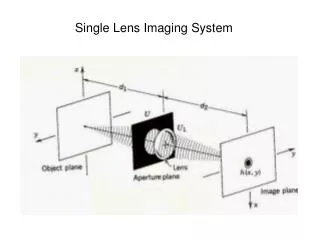
Lens Free Live Cell Imaging Ian Pykett, Vassilios Albanis. The Phase Focus Virtual Lens™. A method for imaging and microscopy that transfers the task of image formation from physical components (lens) to a software algorithm

E N D
Lens Free Live Cell Imaging Ian Pykett, Vassilios Albanis
The Phase Focus Virtual Lens™ • A method for imaging and microscopy that transfers the task of image formation from physical components (lens) to a software algorithm • A platform technology – applicable in principle to the entire electromagnetic spectrum • Initial market focus: optical, electron and X-ray microscopy
Benefits of the Virtual Lens • Benefits applicable at all wavelengths: • “Lensless” – no high performance focussing devices required • Imaging of arbitrarily large fields of view within extended specimens • Post-acquisition multi-focal-plane reconstructions though the specimen thickness • Routine acquisition of quantitative phase data • Resolution limited in principle only by wavelength • The generic product (a processor-embedded Virtual Lens) can be readily interfaced with existing instrumentation
Technology • Hardware Step: • Moving illumination • Creates multiple “diffraction” patterns from the specimen • Software Step: • Iterative phase retrieval algorithm: the Virtual Lens, a.k.a. Ptychographical Iterative Engine (PIE) • From the diffraction patterns, reconstructs two separate images, from any (or every) focal plane within the specimen • Absorption image (traditional “brightfield” image) …how much the specimen absorbs the light • Quantitative phase image …how much the specimen changes the phase of the light
Diffraction Patterns Technology
Technology Diffraction Patterns
Technology • Iterative Phase RetrievalAlgorithm (Virtual Lens) J. M. Rodenburg, A. C. Hurst, A. G. Cullis. Transmission Microscopy Without Lenses for Objects of Unlimited Size”. Ultramicroscopy 107: 227-231, 2007. J. M. Rodenburg, A. C. Hurst, A. G. Cullis, B. R. Dobson, F. Pfieffer, O. Bunk, C. David, K. Jefimovs, I. Johnson. Hard-X-Ray Lensless Imaging of Extended Objects. Phys. Rev. Lttrs. 98:034801-1 – 034801-4 (2007) J. M. Rodenburg, A. M. Maiden. An improved ptychographical phase retrieval algorithm for diffractive imaging. Ultramicroscopy (2009), doi:10.1016/j.ultramic.2009.05.012 (in press)
Technology Eggs: lily anther Fly’s wing Brightness: absorption. Colour: phase
Retrospective focussing • Multi-focal-plane reconstructions from single acquisition
Specimen characterisation 25.4 mm • Thickness measurement Phase image
Specimen characterisation • Quantitative Refractive Index Measurement Specimen Frog’s egg section Preparation Standard microtome Stain None Field of view 2 mm x 2 mm Acquisition Single pass mode Resolution ~10 µm Wavelength 633 nm
Applications: Stand-alone prototype • Initial conventional low magnification “scout scan” defines region of interest for fully-automated diffraction imaging • Illumination currentlyrastered via specimenstage translation • Acquisition timecurrently ~2 s per spot • In future, via (e.g.)galvanometerlaserscannner • Parallel processing option • Currently ~5 s per iteration(500 x 500 array)
Applications: contact lenses • Analysis of contact lenses suspended in hydration solution • Exemplar for hydrogels; biofilms; etc. • Competing methods (conventional microscopy; atomic force microscopy) are destructive, of limited field of view, uninformative, or can be used only with non-hydrated (e.g.: freeze-dried) lenses Absorption (conventional brightfield) image Phase image Phase discontinuity(“phase island”):Demarcation ofhydrophobic area?
Applications: life sciences • Non-contact microscopy of immersed cells • High contrast without stains • Retrospective focussing • Specimen characterisation (refractive index; thickness) • E.g.: Changes in cellular refractive index are early harbingers of apoptosis • Wide field of view (typically 400µ2 – 2mm2) • Field of view independent of resolution • Large working distance(30mm+)
Cell imaging Fibroblasts Absorption image Phase image Cell type Keratinocytes Cell System Single layer adherent Medium Phosphate buffered solution Cell age ~ 2 days Vessel 8-well plate Stain None Working 30mm distance Field of view 1 mm x 1 mm Acquisition Single pass mode Resolution ~1 µm Wavelength 405 nm Keratinocyte colonies
Cell imaging Absorption image Phase image Cell type Human dermal fibroblasts Cell System Single layer adherent Medium Phosphate buffered solution Cell age ~2 days Vessel Sealed 35 mm plastic petri dish Stain None Working 30 mm distance Field of view 1 mm x 1 mm Acquisition Single pass mode Resolution ~1 µm Wavelength 405 nm Absorption (conventional brightfield) image Phase image
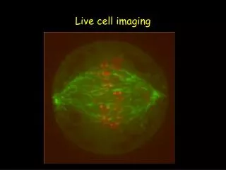
Live cell imaging Absorption image Phase image Cell type Human epithelial Cell System Multilayer adherent Medium Epidermal Growth Factor Cell age 5 - 7 days Vessel T-25 flask Stain None Working 30 mm distance Field of view 1 mm x 1 mm Acquisition Single pass mode Resolution ~1 µm Wavelength 405 nm Absorption (conventional brightfield) image Phase image
Cell segmentation Phase image Cell type Human metastatic melanoma Cell System Single adherent Medium Phosphate buffered solution Cell age 5 - 7 days Vessel T-25 flask Stain None Working 30mm distance Field of view 1 mm x 1 mm Acquisition Single pass mode Resolution ~1µm Wavelength 405 nm Post-processing Matlab application Segmentation Standard threshold technique Segmented image
Cell imaging applications strategy • Confirm the unique combination of benefits of the Virtual Lens for high content screening applications: • Goal: Automated in situ live cell imaging to report on cell death, proliferation, and cell cycle dynamics on a 3D basis over prolonged periods of time
Acknowledgements • University of Sheffield • John Rodenburg(Phase Focus CSO) • Andrew Maiden • Sheila McNeil • Louise Smith • University of York • Peter O’Toole • Cardiff University • Nick White • Rachel Errington • University of Manchester • Sandra Downes • Michael Read