Download
1 / 24
240 likes | 585 Views
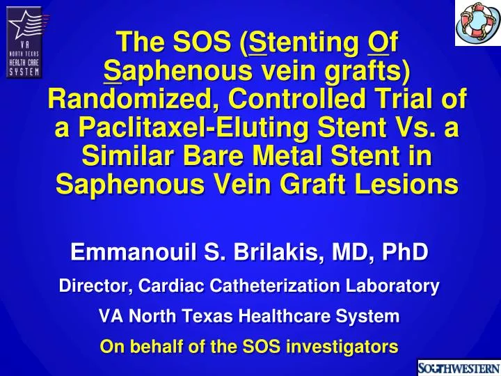
The SOS ( S tenting O f S aphenous vein grafts) Randomized, Controlled Trial of a Paclitaxel -Eluting Stent Vs. a Similar Bare Metal Stent in Saphenous Vein Graft Lesions. Emmanouil S. Brilakis , MD, PhD Director, Cardiac Catheterization Laboratory VA North Texas Healthcare System

E N D
The SOS (StentingOf Saphenous vein grafts) Randomized, Controlled Trial of a Paclitaxel-Eluting Stent Vs. a Similar Bare Metal Stent in Saphenous Vein Graft Lesions Emmanouil S. Brilakis, MD, PhD Director, Cardiac Catheterization Laboratory VA North Texas Healthcare System On behalf of the SOS investigators
I, EmmanouilBrilakis DO NOT have a financial interest/arrangement or affiliation with one or more organizations that could be perceived as a real or apparent conflict of interest in the context of the subject of this presentation. Disclosure Statement of Financial Interest
Veteran Affairs, VISN-17 Clark R. Gregg fund, Harris Methodist Foundation, Fort Worth, Texas Funding
SOS design • DESIGN: Prospective, randomized, multi-center trial comparing the Taxus™ paclitaxel-eluting stent (PES) with a similar Express2™ bare metal stent (BMS) in saphenous vein graft (SVG) lesions • OBJECTIVE: To compare the 12-month angiographic and 24-month clinical outcomes between PES and BMS in SVG lesions • PRINCIPAL INVESTIGATOR: Emmanouil S. Brilakis, MD, PhD. VA North Texas Healthcare System, Dallas, Texas
SOS: sample size determination • 31 patients needed per group to have 80% power (2-sided alpha 0.05) to detect a 66% reduction in binary angiographic restenosis assuming 50% restenosis in BMS group and 1 lesion treated per pt • Target enrollment: 40 patients per group to account for losses during follow-up
SOS: Patient flow 80 patients (112 lesions) enrolled between 2005 and 2007 in 5 clinical sites in USA and Europe BMS 39 pts 55 lesions PES 41 pts 57 lesions Died: 4 pts Emergent CABG: 1 pt Declined: 3 pts Died: 1 pt Declined: 5 pts Angiographic follow-up at 12 months 33 pts 47 lesions Angiographic follow-up at 12 months 33 pts 43 lesions Died: 1 pt Died: 1 pt Clinical follow-up at 24 months Clinical follow-up at 24 months median follow-up: 18 months
SOS trial centers • Onassis Cardiac Surgery Ctr • Athens, Greece • Vassilios Voudris, MD • Panagiotis Karyofillis, MD • Little Rock VAMC • Joe K. Bissett, MD • Rajesh Sachdeva, MD • Iowa City VAMC • James Rossen, MD • VA North Texas HCS • Coordinating Ctr • Emmanouil Brilakis, MD, PhD • Subhash Banerjee, MD • Christopher Lichtenwalter, MD • James de Lemos, MD • Owen Obel, MD • Michele Roesle, RN • Michael E. DeBakeyVAMC • BiswajitKar, MD Collaborators: Peter Berger, MD Panayotis Fasseas, MD
Late Loss BMS (47 lesions), PES (43 lesions) ± 1.03 ±0.98 mm ±0.57 ±0.54 Diff (95% CI) -0.81 (-0.48, -1.46) P<0.0001 Diff (95% CI) -0.87 (-0.51, -1.22) P<0.0001
Binary angiographic restenosis Primary study endpoint % relative risk: 0.18 95% CI: 0.07, 0.48 p < 0.0001
Cumulative frequency distribution curves 100 Before intervention 75 BMS PES % 50 25 0 0 1 2 3 4 Minimum lumen diameter (mm)
Cumulative frequency distribution curves 100 Before intervention 75 BMS PES % 50 After intervention (in-stent minimum lumen diameter) After intervention 25 0 0 1 2 3 4 Minimum lumen diameter (mm)
Cumulative frequency distribution curves 100 75 BMS PES Follow-up BMS % 50 After intervention 25 Follow-up PES 0 0 1 2 3 4 In-stent minimum lumen diameter (mm)
Clinical outcomes median follow-up: 1.5 years
Death from any cause 60 50 Hazard ratio, 1.56 P=0.27 40 % of Patients 30 SBO 20 PES Lung CA Stroke 10 COPD unknown MI MI BMS 0 1.5 0.5 0 1 2 Years from stenting No. at risk BMS 39 37 31 22 12 PES 41 40 34 19 12
Myocardial infarction 60 50 Hazard ratio, 0.67 P=0.10 40 BMS % of Patients 30 20 PES 10 0 1.5 0.5 0 1 2 Years from stenting No. at risk BMS 39 30 23 15 10 PES 41 39 30 15 8
Target lesion revascularization 60 50 Hazard ratio, 0.38 P=0.003 40 BMS % of Patients 30 20 10 PES 0 1.5 0.5 0 1 2 Years from stenting No. at risk BMS 39 33 23 13 8 PES 41 40 32 17 10
Target vessel failure Cardiac death, MI, TVR 60 BMS 50 Hazard ratio, 0.65 P=0.03 40 % of Patients 30 PES 20 10 0 1.5 0.5 0 1 2 Years from stenting No. at risk BMS 39 28 19 11 6 PES 41 38 27 13 7
Clopidogrel use P=NS % of Patients
In saphenous vein graft lesions, compared to a similar bare metal stent, the Taxus™ PES resulted in: Significant reduction in 12-month binary angiographic restenosis, target lesion revascularization and target vessel failure Trends for lower target vessel revascularization, myocardial infarction No difference in mortality and stent thrombosis Conclusions