Download
1 / 69
710 likes | 931 Views
Anemia due to Impaired Iron Metabolism. Wu Chunmei.

E N D
Anemia due to Impaired Iron Metabolism Wu Chunmei
This group disorders are caused by impaired iron metabolism which include:(a) Iron deficiency anemia: deficiency of iron(b) Sideroblastic anemia:`impaired utilization of iron(c) Anemia of chronic disease: defective iron reutilization
The total body iron varies from 3 to 4 g, depending on the sex and weight of the individual. It is greater in males than in females and it increases roughly in proportion to body weight.Male: 50-55mg/KgFemale: 35-40mg/Kg Amount and distribution: 1ml blood=0.5mg Fe 1gHb=3.3mgFe 1mlRBC=1~2mgFe
Compartments iron content(mg) total body iron(TBI)Hb iron: 2670 60-70% Tissue ironmyoglobin 140 3.3%Labile iron pool 80-90 2.2%cytochromes 8 0.2% catalase peroxidaseStorage (available) ironFerritin 440 30% Haemosiderin 390Transport iron 3 0.1% The iron is distributed in several forms:
Absorption ----excretion Balance of iron metabolism
Absorption Transport of iron Utilization Affecting factors?
absorption Duodenum and upper jejunum Fe 3+ in food combined receptor reductase Epithelial cells brush border apo-Ferritin + Fe 3+ Fe 2+ ferritin Fe 2+ Free Fe 3+ stomach reduced(VitC, GSH) gastric juice Fe 2+ Circulation Fe 2+ Tf + Fe 3+ Tf-Fe 3+ Trans-port liver Iron cycle Storage inMMS FerritinHaemosiderin Normablasts Ret. marrow for Hb In tissue: myoglobin heme-containing enzymes
Transport of iron transferrin Serum beta-globulin that binds and transports iron transferrin receptors
apoferritin + Fe MMS Ferritin: An iron-containing protein complex that is formed by a combination of ferric iron with the protein.(apoferritin) Haemosiderin: insoluble storage iron, golden yellow or brown granules in unstained tissue; blue granules when stained with potassium ferrocyanide. It contains more iron than ferritin and aggregates into granules, microscopically visible in tissue and phagocytes. Storage of iron
protophorphyrin + Fe 2+ Heme SA Fe 3+ Tf-Fe 3+ Tf 80% 1/3 Fe 3+ Fe 2+ Ferritin Heme+globin Hb sideroblasts Iron utilized in normoblast
Old RBC blood Hemosiderin ferritin apo-Ferritin Hb globin heme Fe 3+ Fe 2+ Normoblasts Hepatocytes Placental cells have more receptors Storage iron in macrophages Reutilization of Iron
IRON DEFICIENCY ANEMIA (IDA)Iron deficiencyis the state in which the content of iron in the body is less than normal. When the supply of iron to the marrow is insufficient for the requirements of Hb synthesis, IDA develops with varying degrees of microcytic hypochromic anemia.
Causes of iron deficiency : A: Decreased iron intake--Poor diet--Impaired absorption B: Increase iron loss (1ml blood =0.5mgFe)--losses from gastrointestinal tract--Neoplasm--Peptic ulcer--Others (hookworm disease)--Menometrorrhagia --Losses from urine (PNH)--Losses from sputum (rare) C: Increased requirements--Early childhood and adolescence--Women during the reproductive years
Pathogenesis of ID --Lack of iron interferes with heme synthesis, which leads to reduced Hb synthesis and defective erythropoiesis. --There is decreased activity of iron-containing proteins. --Neurologic dysfunction may occur, with impaired intellectual performance, paresthesias, etc. --Gastric acid secretion is reduced, often irreversibly. --Atrophy of oral and gastrointestinal mucosa may occur
Clinical Features 1. General symptom of anemia:Fatigue,weakness,or palpitations, headache 2. Essential iron deficiency: --Children may have poor attention span, poor response to sensory stimuli, retarded developmental and behavioral achievement, irritability and retarded longitudinal growth. --Paresthesias and burning of tongue may occur. --Pica, craving to eat unusual substances such as clay,or ice, is a classic manifestation.
Physical examination Pallor Smooth red tongue, stomatitis Angular cheilitis Koilonychia(rare) Retinal hemorrhages/exudates(severe anemia) Accelerated retinopathy in diabetics Splenomegaly(occasionally)
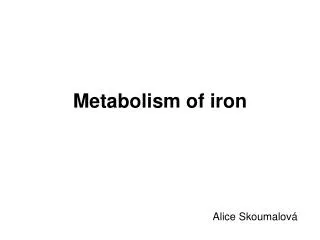
Laboratory Findings 1.Blood: Hypochromic microcytic anemia • RBC↓,Hct ↓, Hb ↓:male<120g/l, female<110g/l, pregnant women<100g/l, MCV<80gl, MCH<26g, MCHC<31% • RDW ↑(>14%, earliest change ) • morphology: anisocytosis , mild ovalocytosis, target cells, ring cells, elongated hypochromic elliptocytes (pencil cells), nucleated RBC, basophilic stippling RBC • Ret normal or reduced • Leukocyte normal or decreased • Platelets increased or decreased
IDA早期血象:红细胞生成缺铁期,部分红细胞生理性中心浅染区轻度扩大,临床上无贫血IDA早期血象:红细胞生成缺铁期,部分红细胞生理性中心浅染区轻度扩大,临床上无贫血
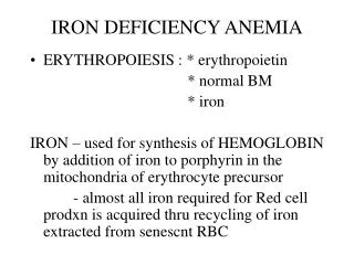
2. Marrow: hypercellularity anemia • marrow usually hypercellularity with M/E ratio variable. • morphology: erythroid hyperplasia and mainly late normoblasts, which may be small, with narrow rim of ragged cytoplasm and poor Hb formation (polychromatic) and densed nucleus(old nucleated young cytoplasm). Basophiloic stippling and Howell-Jolly body may present in normoblasts.The mature RBCs as in blood.
IDA骨髓象:中幼红细胞、晚幼红细胞增生为主,胞体较小,胞浆边缘不规则呈锯齿状、胞浆偏蓝,呈“幼浆老核”现象。IDA骨髓象:中幼红细胞、晚幼红细胞增生为主,胞体较小,胞浆边缘不规则呈锯齿状、胞浆偏蓝,呈“幼浆老核”现象。
IDA HA
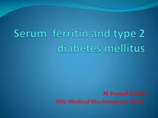
3 The indexes of iron metaolism • ①Marrow stainable iron. • Reference value: • -extracellular iron (+~++) • -intracellular iron (19%~44%) It is a direct and reliable index to reflect the level of storage iron. It decrease (<15%),or absent in ID Sideroblasts decrease in IDE
正常:内铁阳性 IDA骨髓铁染色:内铁阴性
② SF(serum ferritin)and EF(erythrocyte alkaline ferritin) SF Reference value: --< 10µg/l in IDA --10 ~ 20µg/l are presumptive, but not diagnostic. --May be elevated with concomitant inflammation diseases . IDA can be suspected in rheumatoid arthritis if SF is less than 60µg/l or less than 30µg/l in chronic inflammation. Adult male:50~200µg/l; It is a sensitive and reliable index to evaluate total body iron stores, but can be interfered by some conditions. EF reference value: less sensitive than SF, <6.5µg/E in IDA
③Serum iron concentration SI is a direct measure of the amount of iron bound to transferrin. Reference value:50-150µg/dl Adult M:11.6~31.3µmol/l; F:9.0~30.4µmol/l(A:20µmol/l) transporting iron, with a lot of affect factors
④TIBC: total iron binding capacity TIBC is a measure of the amount of iron that can be bound by transferrin. Reference value: 360~390µg/l TIBC:male:50~77µmol/l; female 54~77µmol/l • Usually increased in ID. • decreased in liver disease, malignant tumor, HA, chronic renal disease…
⑤TS (Transferrin Saturation): SI TS = ×100% , TIBC In normal condition, 1/3 transferrin binds to iron. Reference value: 20~50%. ( A:30%) 15% or less in patients with IDA ; >50~60% resulting in iron loading.
UIBC SI TIBC
⑥sTfR(serum soluble transferrin receptor) : Reference value: 5~9μg/L(ELISA) sTfR level increase in IDE, IDA (when iron store are exhausted) sTfR level also increase in other disease with ineffective or effective erythroid precursor proliferation
⑦ FEP (Free Erythrocyte Protophorphyrin) andZPP( protophorphyrin binds to zinc) --usually increased in ID --Very sensitive for diagnosis of ID and suitable for large-scale screening of children , detecting both ID and lead poisoning.(why?) FEP less sensitive than SF and EF.
TIBC TS SI Sensitivity of indexes of iron: SF Marrow stainable iron EF sTfR FEP
Diagnosis of IDA: ①+more than two of ②~⑧ ①Hypochromic microcytic anemia(Hb male < 120 g/l , female<110g/l, pregnant women<100g/l, MCV < 80fl, MCH<26g, MCHC < 31% , RDW > 14% , and morphologic changes) ② Identified causes associated with ID and significant clinical symptoms ③ SI <10.7µmol/L,and TIBC>64.44 µmol/L ④TS<0.15 ⑤ Extracellular iron (-), sideroblasts <15% or absent ⑥ FEP>0.9 µmol/L(blood) ,or ZPP>0.96umol/L(blood), or FEP/Hb >4.5µg/gHb ⑦ SF<14µg/L; ⑧Effective to therapy with iron
1. Iron depletion (ID): It is the earliest stage of iron deficiency. In which storage iron is decreased or absent but serum iron concentration and blood Hb levels are normal. ---Extracellular iron is decreased or absent ---SF concentration falls There are three stages of iron deficiency:
2.Iron deficiency erythropoiesis (IDE): It is a somewhat more advanced stage of iron deficiency. In which deficit of the functional iron compartment is associated with the development of iron deficiency erythropoiesis. It is characterized by decreased or absent storage iron, usually low SI and transferrin saturation, without frank anemia.
manifestations --Extracellular iron is absent--Intracellular iron is decreased--Serum soluble transferrin receptor(sTfR) is increased.--TIBC is increased--Serum iron level falls--Transferrin saturation falls--An increase in the RDW--generally asymptoms.
3.Iron deficiency anemia (IDA): It is a most advanced stage of iron deficiency. It is characterized by decreased or absent iron stores, low SI , low transferrin saturation and low Hb or hematocrit value. Besides the above characteristics, there are:--red cell count decrease --Many symptoms
Diagnosis process of -Anemia? -Microcytic hypochromic anemia? -IDA?(measurements of iron metabolism) -The cause of IDA!
Sideroblastic Anemias (SA) The sideroblastic anemias are a heterogeneous group of disordes that have as common features the presence of large number of ringed sideroblasts in the marrow, ineffective erythropoiesis, increased levels of tissue iron and varying proportions of hypochronic erythrocytes in the blood.
hereditary:x chromosome-linked ,partially recessive inheritance, males are anemic and females are carriers.autosomally-linked, or mitochondria entitiesacquired :primary:neoplasia (MDS-RAS) secondary: drugs, toxin, alcohol, or coincident to neoplastic or inflammatory disease Classification: