Download
1 / 44

E N D
MICROBIAL CAUSES OF BONE INFECTION AND ARTHRITIS 1
Objective of the lecture At the end of the lecture, students must be able to: • Describe the causative agents of osteomyelitis & septic arthritis • Identify the mechanisms of transmission of pathogens in osteomyelitis & septic arthritis • Discuss the types of osteomyelitis & septic arthritis • Describe the key risk factors for osteomyelitis & septic arthritis • Describe diagnosis of osteomyelitis & septic arthritis 2
1. BONE INFECTIONS Bone & joint infections are serious - may be life- threatening, lead to long-term disability & reduced quality of life. Infections may exist separately or together. Both are common in infancy & childhood. Infections can cause growth impairments in children - when epiphysis is involved. Types of bone & joint infections: 4 main diseases 1. Osteomyelitis 2. Septic Arthritis 3. Prosthetic joint infections 4. Reactive arthritis – Immunological following infections 3
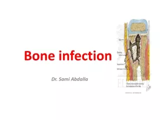
OSTEOMYELITIS • Osteomyelitis: inflammation of the bone, bone-marrow & surrounding soft tissue. • It is a progressive infection that results in inflammatory destruction followed by new bone formation. • Nelaton credited with introducing the term osteomyelitis in 1844. • Osteomyelitis is most common in children of 3-12 year • In children most often the consequence of bacteremia. • Most osteomyelitis are caused by staphylococci, commonly found on the skin or in the nose of even healthy individuals. 4
Osteomyelitis Classification based on 1. Duration: – Acute osteomyelitis – Subacute osteomyelitis – Chronic osteomyelitis. 2. The type of Host response to the infection: • Pyogenic: Acute & chronic osteomyelitis • Non-Pyogenic - Granulomatous: TB, Syphilis, Fungal, etc 3. Mechanism: – Exogenous – Hematogenous 5
Osteomyelitis 1. Acute osteomyelitis • Pyogenic infection of <2 weeks in duration - Symptoms present within 2 weeks after infection • Children have acute osteomyelitis form more often (60-70%) than adults do, at a rate of about 1:5000. Acute hematogenous osteomyelitis (AHO) • Most common type of bone infection, usually seen in children 6
Osteomyelitis Etiology of hematogeneous osteomyelitis Acute hematogenous osteomyelitis (AHO) 1.Children: – S. aureus (60-90%) – Group B Streptococci (Streptococcus agalactiae) – H. influenzae: children 6 months to 4 years old – E. coli (in neonates) – S. pneumoniae – Kingella kingae are also common – Salmonella (Children with sickle cell disease) 7
Osteomyelitis 2. Adults: – S. aureus (55%) – E. coli – Klebsiella – Pseudomonas – in patients with GUT infections or with IV drug abusers – Streptococci – Actinomyces israeli (mandible) 8
Acute osteomyelitis - Primary site of infection usually in the metaphysial region. 9
Osteomyelitis 2. Subacute Osteomyelitis • More insidious onset and lacks severity of symptoms • Duration: 2-6 weeks • Subacute osteomyelitis most commonly affects the metaphysis, followed by the diaphysis, and rarely the epiphysis. 14
Osteomyelitis Its relative mildness is due to: a. Organism being less virulent OR b. Patient more resistant OR c. Both Causative organisms Subacute Osteomyelitis • S. aureus (30-60%) & S. epidermidis are predominant organisms identified in subacute osteomyelitis • Streptococcus • H. influenzae • P. aeruginosa (IV drug user) • Salmonella (in sickle cell anemia) • K. kingae. 16
Chronic osteomyelitis 3. Chronic osteomyelitis • Duration: >6 weeks; in children is uncommon. Types of chronic Osteomyelitis a. Complication of acute osteomyelitis b. Post-traumatic (Secondary to open fractures) c. Post operative Chronic suppurative osteomyelitis • Common in mandible, associated with odontogenic infections. • Polymicrobial infection, predominating anaerobes such as Bacteriods, Porphyromonas or Provetella. 12
Chronic osteomyelitis Etiology • Primary resulting from low virulent microorganisms. • Secondary sequelae of acute suppurative osteomyelitis • S. aureus (commonest cause). • S. epidermidis (commonest in surgical implant) • S. pyogenes • E. coli • P. aeruginosa – form biofilm • Salmonella spp, S. aureus cause osteomyelitis in patients with sickle cell disease – • Serratia marcescens & E. coli - isolated from contiguous infection in another part of the body, E.g: Sinus, Ear, Dental, Respiratory & GU infections. 13
Chronic osteomyelitis • Laboratory: – Bone biopsy is essential for diagnosis – Microbiological cultures for bacteria, mycobacteria & fungus are required for appropriate treatment 14
Granulomatous Osteomyelitis Non-pyogenic (granulomatous) causes of osteomyelitis 1. Tuberculous osteomyelitis 2. Congenital/acquired syphilis (skeletal syphilis) 3. Actinomycotic (Myecetomal) osteomyelitis 4. Fungal osteomyelitis: Histoplasma, blastomycosis, coccidioidomycosis 5. Parasitic infestation - e.g. hydatid. 15
Granulomatous Osteomyelitis Spread in chronic osteomyelitis 1. Hematogenous seeding: • Usually monomicrobial, occurs in children <12 years of age although adults can have this disease 2. Contiguous or exogenous • From adjacent soft tissues & joints: E.g. wounds, abscess …. Usually polymicrobial. • Organisms: S. aureus (70%- 80%), Pseudomonas 3. Direct Inoculation Osteomyelitis • As a result of trauma, surgery; Usually polymicrobial • S. aureus: the most common causative organism. 16
TB Osteomyelitis Skeletal TB • Dissemination of TB outside the lungs - skeletal TB. • TB osteomyelitis is secondary to hematogenous spread from a primary source in the lung or GI tract. • The spinal column is involved in less than 1% of all cases of TB • Most common in children & young adults • It commonly occurs in the vertebrae & long bones. • It involves mainly vertebrae / thoracic & lumbar/(Pott disease, also known as tuberculous spondylitis) followed by long bones - knee & hip. 17
Syphilitic osteomyelitis 1. Congenital syphilis • Caused by Treponema pallidum, STD. • Transplacental spread of TP from mother to fetus • Hematogenous spread such as the tibia, are mainly affected, cause 2 chief bone lesions: Periosteitis & Osteochonditis 2. Acquired syphilis • Bone lesions are manifestations of tertiary syphilis. • Gummatous lesions appear as discrete punched-out radiolucent lesions in medulla or destructive lesions within the cortex. The surrounding bone is sclerotic & with no discharge • Bones frequently affected: Nose, palate, skull, extremities, esp. the long tubular bones such as tibia of T. pallidum - Long bones, 18
Fungal Osteomyelitis • Bone fungal infection is rare but potentially life threatening condition • Usually reported in immunocompromised hosts • E.g. Maduromycoses Treatment • No effective chemotherapy – – Amphoterecin B - IV used, but toxic – Wide excision – amputation 19
Actinomycotic osteomyelitis Actinomyces israelii • Filamentous, Gram-positive, non–acid-fast, anaerobic- to-microaerophilic bacteria. • Actinomycosis is subacute-to-chronic granulomatous disease. • Two-thirds of all cases occur in the cervicofacial region • Bacteria found as normal flora of the oral cavity • Remain localized in the soft tissues or invade the jaw bones. • The more aggressive lesion resembles chronic suppurative osteomyelitis 20
Osteomyelitis Diagnostic approaches • Physical exam: bone tenderness, swelling, redness • Microbial etiology confirmed in 75% of osteomyelitis Tests may include: – Blood culture: positive in about 50% of cases – Bone biopsy - Needle aspiration of the area around affected bones for culture, staining or histologic exam – Bone X-ray: in later stage – demineralization seen – MRI or CT scanning – CBC for Leukocytosis – Elevated C-reactive protein, Elevated ESR 21
2. JOINT INFECTIONS Septic Arthritis 22
Septic Arthritis (SA) Septic arthritis (Infectious arthritis): • Inflammation of joint space due to bacteria, fungi, viruses or parasitic infections. • It is inflammation of a synovial membrane with purulent effusion into the joint capsule. • Septic arthritis is a painful infection in a joint that can come from mos that travel through bloodstream from another part of your body. Septic arthritis can also occur when a penetrating injury, such as an animal bite or trauma, delivers mos directly into the joint. • Infants & older adults are most likely to develop septic arthritis. 23
Septic Arthritis (SA) Etiology of septic arthritis • Bacteria are the most significant pathogens in septic arthritis because of their rapidly destructive nature. S. aureus – the most common: 60-80% of cases. Streptococcus – 2ndcommon (GAS, S. viridans, S. pneumoniae, GBS): 20% 24
Septic arthritis N. gonorrhoeae: 75% among young sexually active individuals, 3x in women than men. Gram negative rods: E. coli in elderly, In IV drug users - Pseudomonas Other bacterial causes: N. meningitidis, Borrelia burgdorferi, Mycobacterium tuberculosis, Staph. epidermidis → Prosthetic joints; Salmonella spp in individuals with SLE Brucella, mycoplasma/ureaplasma Kingella kingae -has emerged as a major cause of septic arthritis in young children. 25
Fungal agents: histoplasma, sporothrix schnkii, cocidioides imitis, blastomyces Viruses:HBV, Rubella virus 26
Septic arthritis Pathogenesis & transmission • Septic arthritis develops when bacteria or other mos gain entrance to a joint via 3 routes: 1. Hematogenous spread – common in IV drug injection 2. Direct inoculation – Injury, intra-articular injection, arthroscopy or orthopedic surgery especially insertion of joint prosthesis. 3. Contiguous spread from adjacent focal infections - (e.g. penetrating trauma). More common in children. • Extension of osteomyelitis through epiphysis or by lateral extension through periosteum into joint capsules. 27
Septic arthritis • Soft tissue infections: cellulitis, abscess, bursitis, tenosynovitis. • S. aureus has a variety of receptors, termed microbial surface components recognizing adhesive matrix molecules for host proteins that mediate adherence to the joint extracellular matrix or implanted medical devices. • Some of the host matrix proteins include fibronectin & laminin (adherence proteins), elastin (imparts elastic properties), collagen (structural support) & hyaluronic acid (a glycosaminoglycan that is rich in the joints & the matrix and provides cushioning through hydration of its polysaccharides). 28
Risk factors for septic arthritis 1. Age >80 years 2. Chronic illness (Cancer, RheumatoidArthritis, SLE, Diabetes Mellitus, HIV infection) 3. Prosthetic joint - Hip or knee prosthesis 4. Previous arthritis 5. Recent joint surgery 6. Endocarditis or Bacteremia 7. Skin infection 8. IV drug abuse 9. Alcoholism 10.Trauma 11.Sickle cell anemia 29
Acute Septic arthritis 1. Acute bacterial septic arthritis • Acute septic arthritis is a joint infection that evolves over hours or days. • Synovial membrane is highly vascularized • Acute monoarthritis is septic until proven otherwise. • Cartilage in joints become damaged within hrs or days. • It can affect healthy people and people at high risk. It may develop as a result of: 1. Hematogenous seeding 2. Direct introduction 3. Extension from contiguous focus of infection 30
Acute Septic arthritis Causative agents • S. aureus (50%) - Any age • Streptococcus spp: Streptococcus viridans, S. pneumoniae & Group B streptocci • GNB (10%) - E. coli & pseudomonas common • Neonates: S. aureus, GBS, GNB (E. coli, Proteus, Klebsiella, Pseudomonas) • 1 month- 4 yrs: H. influenzae (children <2 yrs), S. aureus, S. pyogenes, S. pneumoniae, N. meningitidis • 4-16 years: S. aureus • 16-40 years:- N. gonorrhoea, S. aureus • >40 years:- S. aureus 31
Chronic Septic arthritis 2. Chronic septic arthritis Less common: about 5% of infectious arthritis; Often due to fungal, mycobacterial & viral affect only one joint or, occasionally, several joints. Most often affects people who are at high risk. Most commonly infected joints include the knee, wrist, hip, shoulder, elbow and joints in the fingers. 32
Chronic Septic arthritis Etiology • Mycobacterium tuberculosis -TB • Borrelia burgdorferi - Lyme disease • Treponema pallidum - Syphilis • Mycoplasmas: M. pneumoniae, M. hominis • Fungi: C. albicans in AIDS cases; other • Viruses: HBV, Rubella, Mumps, Varicella,Adenovirus, Coxachie, Parvoviruses, EBV 33
Diagnosis of acute & chronic septic arthritis Diagnosis • Analysis of synovial fluid for cell count, protein, glucose, exam of crystals under microscope This fluid will show: – Presence of microorganisms – Presence of Ab directed against the mos – Turbidity – More than 10,000 pmns/mm3 – Decreased glucose conc. (<0.6% of blood glucose) – Increased lactic acid concentration (> 65 mg/dl) 34
Diagnosis of Septic arthritis • Gram stain & culture, chemical analysis. – Blood culture (positive in 30-60%): at least 2 sets of blood cultures to R/o a bacteremic origin of the septic joint – Microbial etiology confirmed in ⅔ of cases of septic joint. – Histologic exam of tissues in granulomatous forms. – In chronic arthritis synovial biopsy may distinguish b/n an septic and a non-infection process. – Serology for Lyme disease, Brucellosis 35
Diagnosis of Septic arthritis Other tests – PCR: Molecular methods to detect bacterial DNA in synovial fluid, e.g. B. burgdorferi, M. hominis, N. gonorroheae. – X-ray of affected joint 36
Prosthetic Joint Infections • Prosthetic joint infection (PJI): infection involving the joint prosthesis & adjacent tissue. • Between 1-5% of all prosthetic joints become infected and is one of the leading causes of arthroplasty failure. • 98% are hips & knees, the remainder mostly shoulder. • PJI can be acute (<4 weeks) or chronic Etiology of Infection 1.Early onset (<3 months): – Organisms gain entry at the time of operation – Organisms: S. aureus, Coliforms, mixed infections 37
Prosthetic Joint Infections 2. Delayed (3–24 months): – Coagulase negative Staphylococci, Propionibacterium spp 3. Late onset (>24 months): • Spread from a distant source of infection • S. aureus, E. coli, Coliforms • 50% have no apparent source 38
Prosthetic Joint Infections Summary of etiologic agents 1.Gram positive: 65% • Gram positive – CONS > S. aureus > Streptococcus > Enterococci a. Coagulase negative staphylococci (CoNS): 22% b. S. aurues: 20% c. Streptococci: 14% d. Enterococci: 7% 1. G-ve cocci: 25% • Enteric > pseudomonas 2. Anaerobes: least common - 10% 39
Prosthetic Joint Infections Pathogenesis • Locally introduced (60-80%) – Operative contamination – Wound sepsis contiguous to the prosthesis • Haematogenous (20-40%) – Any bacteremic episode may seed a prosthetic joint – S. aureus bacteremia leads to 34% incidence of PJI • Biofilms: populations of mos adhering to environmental surfaces & bioprosthetic materials. • These mos are usually encased in an extracellular polysaccharide that they themselves synthesize. 40
Prosthetic Joint Infections Risk factors Artificial joint implants • Bacterial infection somewhere else in the body • Chronic disease (DM, RA, sickle cell disease) • IV drug use • Immunocompromised states • Recent joint injury • Recent joint arthroscopy or surgery • Poor nutritional status • Obesity • Advanced age • Smoking 41
Prosthetic Joint Infections In Children • Occurs most often in those younger than 3 years. • The hip is often the site of infection in infants. • Organisms: Group B Streptococci or H. influenzae (if not vaccinated). Presenting symptoms • Joint pain: 95% • Fever: 43% • Periarticular swelling:38% • Wound / sinus drainage: 32% 42
Prosthetic Joint Infections Diagnosis: • Radiography: changes - Loosening of prosthesis • Arthrocentesis: Synovial fluid leukocyte count >1700/mm3 • Histopathology: Peri-prosthetic tissue Management • Goal of treating infection: pain-free, functional joint 1. Surgical • Debridement with retention of prosthesis • Implant removal 43
ThankYou 44