Download
1 / 8
80 likes | 140 Views
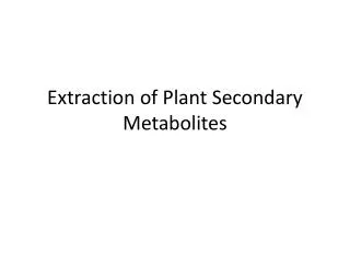
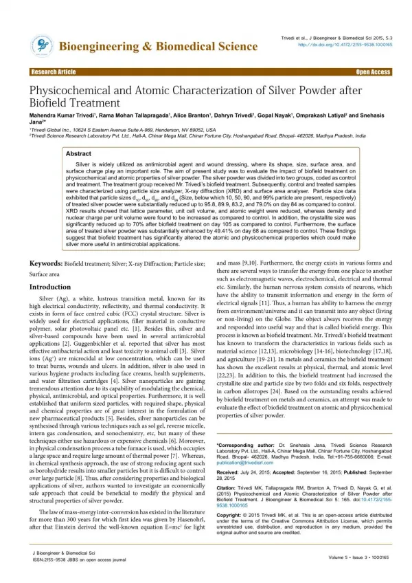
Digera muricata (L.) Mart (Amaranthaceae), commonly known as ‘Latmahuria’ is traditionally used medicinal plant. Laboratory evolutions were made to assess the quantification of primary<br>metabolites in Digera muricata. It contains higher soluble sugars in roots, starch, protein and lipids in leaves and phenols in roots as compared to other parts of the plant. Results indicate that methanolic extracts of plant parts showed significant antimicrobial activity. The leaf extract was<br>found to have maximum activity index against Escherichia coli and Fusarium oxysporum when tested by the disc diffusion method. Aspergillus niger showed significant activity against root extract. <br>

E N D
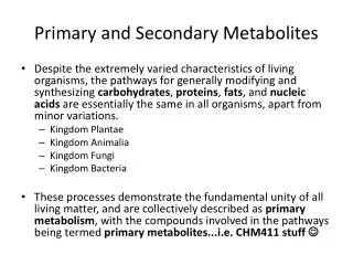
Available on line www.jocpr.com Journal of Chemical and Pharmaceutical Research __________________________________________________ J. Chem. Pharm. Res., 2011, 3(2):424-431 ISSN No: 0975-7384 CODEN(USA): JCPRC5 Study of primary metabolites and antimicrobial activities of Digera muricata (L.) Mart Neha Sharma*, Babeet Singh Tanwer and Rekha Vijayvergia Plant Pathology and Plant Biochemistry Laboratory, Department of Botany, University of Rajasthan, Jaipur, India ______________________________________________________________________________ ABSTRACT Digera muricata (L.) Mart (Amaranthaceae), commonly known as ‘Latmahuria’ is traditionally used medicinal plant. Laboratory evolutions were made to assess the quantification of primary metabolites in Digera muricata. It contains higher soluble sugars in roots, starch, protein and lipids in leaves and phenols in roots as compared to other parts of the plant. Results indicate that methanolic extracts of plant parts showed significant antimicrobial activity. The leaf extract was found to have maximum activity index against Escherichia coli and Fusarium oxysporum when tested by the disc diffusion method. Aspergillus niger showed significant activity against root extract. Keywords;Digera muricata, disc diffusion, primary metabolites, activity index, antimicrobial activity. ______________________________________________________________________________ INTRODUCTION With the development of antimicrobials, microorganisms have adapted and become resistant to previous antimicrobial agents. The old antimicrobial technology was based either on poisons or heavy metals, which may not have killed the microbe completely, allowing the microbe survive, change, and become resistant to the poisons and/or heavy metals. Modern Phytomedicine is a timely and original handbook paving the way to success in plant-based drug development, systematically addressing the issues facing a pharmaceutical scientist who wants to turn a plant compound into a safe and effective drug. Plant derived antimicrobial agents have been largely 424
Neha Sharma et al J. Chem. Pharm. Res., 2011, 3(2):424-431 ______________________________________________________________________________ overlooked. Hence, the Digera muricata (L.) Martis used in both folk and traditional system of medicine. Digera muricata (L.) Mart (Amaranthaceae) wild edible plant commonly known as Lesua, it is also known as False Amaranth, Latmahuria, Kunanjara and Aranya. This is an annual herb, growing to 20-70cm tall. Leaf stalks are long, up to 5cm, base is narrowed, and the tip pointed. Flowers are borne on slender spike-like racemes, which can be as large as 30 cm long. Flowers are hairless, white mixed with pink to carmine or red, usually becoming greenish-white in fruit. Flowering occurs in month of August and September.It is widely distributed in Rajasthan, Maharashtra and Andrapradesh. Leaves and young shoots of D. muricata are locally used as a vegetable and given to relieve constipation [1].Itis also used internally against digestive system disorders and in India flowers and seeds are used to treat urinary discharges [2,3]. Leaf paste is applied locally to prevent pus formation [4]. Boiled root infusion given to mother after child birth for lactation purpose [5]. However no studies of primary metabolites and antimicrobial have been reported on Digera muricata. MATERIALS AND METHODS Plant material: Healthy plants of Digera muricata were collected from University of Rajasthan campus and authenticated by Herbarium, University of Rajasthan, Jaipur, Rajasthan, India. Chemicals: All the chemicals and growth regulators were used are analytical grade and purchased from Hi Media Pvt. Ltd., Mumbai, India. Primary metabolite estimation Extraction of carbohydrates: (A) Total soluble sugars: The dried and milled test sample 50 mg each was macerated in a grinder with 20 ml of ethanol and left for 12 hrs. and mixtures was centrifuged (1200 rpm) for 15 min, the supernatants were removed and was concentrated on a water-bath. The volume of these aqueous concentrates was raised to 50 ml with distilled water (Ext. A) and processed further by following the method of Loomis and Shull [6] for soluble sugars. However, the residual pellet obtained by centrifugation was used for the estimation of starch. (B) Starch: The above residue of each test sample was suspended in a mixture of 5 ml of 52% perchloric acid solution and 6.5 ml of distilled water, shaken vigorously (5 min) and centrifuged (2500 rpm). This step was repeated three times and the supernatants of each sample were pooled and the volume was raised to 100 ml with distilled water (Ext B). Out of this (Ext. B), 1 ml aliquot was taken separately to estimate starch quantitatively [7]. Quantification of carbohydrates: Aliquot (1ml) of each of the test sample from Ext. A and B were used to quantifying the total levels of carbohydrates using phenols-sulphuric acid method [8]. A regression curve for standard sugar (glucose) was also prepared. A stock solution of glucose (100 µg/ ml) was prepared in distilled water, out of which 0.1 to 0.9 ml was transferred to test tube and the volume was raised 425
Neha Sharma et al J. Chem. Pharm. Res., 2011, 3(2):424-431 ______________________________________________________________________________ to 1 ml with distilled water. To each of these, 1 ml of 5% aqueous phenol was added rapidly having kept in an ice chest and shaken gently. Later 5 ml of Conc. H2SO4 was rapidly added by agitating gently during the addition of the acid subsequently, the tube was kept on a water-bath (26º– 30ºC) for 20 min, and the optical density (ODs) of the yellow orange colors thus developed were taken at 490 nm in a Spectrophotometer after having set it for 100% transmission against the blank. Four replicates of each sample were run and there mean values were calculated. A regression was computed between its known concentrations and their respective ODs. This was based on Beer’s Law. The concentration (mg/gdw) of the total soluble sugars was directly worked out from the regression curve of the standard glucose. Four replicates of each experimental sample were taken and their mean values recorded. The sugar content in terms of glucose equivalent and the use of conversion factor (0.9 to convert the values of glucose to starch) was made in each case. Extraction of Proteins: A 60 mg of the dried test sample was macerated [9] in 10 ml of cold TCA (10%) for 30 min kept at low temperature 4º C for 24 hr and then centrifuged. Each of the supernatants was discarded and the resultant pellet was re-suspended in 5% TCA (10 ml) and heated on a water bath at 80º C for 30 min. Each of these samples was cooled, re-centrifuged and each time the supernatant discarded. Later the pellet was washed with distilled water, centrifuged and each of the residues was dissolved in 1N NaOH (10 ml) and left overnight at room temperature. Quantification of Proteins: In each of 1 ml extract, total protein content was estimated using the protocol of Lowry et al [10]. A stock solution (1mg/ml) of bovine serum albumin (Sigma Chemicals) was prepared in 1 N NaOH, from which 0.1 to 0.9 ml of the solution was dispensed separately in a test tube. After this, the volume of each was raised to 1 ml by adding distilled water. To each test sample, 5ml of freshly prepared alkaline solution (prepared by mixing 50 ml of 2% Na2CO3 in 0.1 N NaOH and 1 ml of 0.5%CuSO4. 5H2O in 1% sodium potassium tartrate) was added at room temperature and left undisturbed for a period of 10 min. Subsequently, to each of these mixture tubes 0.5 ml of Folin-Ciocalteau reagent (CSIR centre for Bio-chemicals, Delhi: diluted with equal volume of distilled water just before use) was rapidly added and after half an hr, the OD of each was measured at 750 nm using a spectrophotometer against the blank. Three replicates of each concentration were taken and there mean values were used to compute a regression curve. The total protein content in each sample was calculated by referring the ODs of test sample with the standard curve of BSA. Three replicates were examined in each case and their mean values were recorded. Extraction of Lipids: One g of each of the dried and milled test sample was macerated with 10ml distilled water [11]. To this, 30 ml of chloroform-methanol (2: 1, v/v) was added and mixed thoroughly. Each mixture was left overnight at room temperature; 20 ml of chloroform and the equal volume of distilled water was added and centrifuged. 426
Neha Sharma et al J. Chem. Pharm. Res., 2011, 3(2):424-431 ______________________________________________________________________________ Out of the three layers, a clear lower layer of chloroform containing all lipids was collected in pre-weighted beaker, the solvent evaporated completely and weighed, which was taken as the weight of total lipids/g of the dried tissue sample. Extraction of Phenols: Each of 200 mg dried and milled test samples was homogenized in 80% ethanol (10 ml) for 2 hrs and left over night at room temperature. It was centrifuged, the supernatants were collected individually and the volume of each was raised to 40 ml with 80% ethanol. Quantification of Phenol: To estimate total phenols in each of the test sample, the protocol of Bray and Thorpe [12] was followed, wherein a standard curve of caffeic acid (a phenol) was prepared A stock solution (100 µg/ml) of caffeic acid was prepared in 80% ethanol, from which 0.1 to 0.9 ml was transferred into test-tubes separately and the volume in each case was raised to 1 ml with 80% ethanol. To each of these tubes, 1 ml of Folin–Ciocalteau reagent (prepared by diluting the reagent with distilled water in 1:2 ratio just before use) accompanied by 2 ml of 20% Na2CO3 solution was added and the mixture was shaken vigorously. Each of these were boiled on a water bath (1 min), cooled and diluted to 25 ml with distilled water. The OD was taken at 750 nm using a spectrophotometer against a blank. Three such replicates were taken for each concentration and the average OD was plotted against the respective concentration to compute a regression curve. Each test sample was processed in this similar manner, ODs were measured and the total level of phenols was calculated from the mean values (with reference to caffeic acid) by referring the OD of the test sample with the regression curve of the standard. Preparation of extracts: The stem, leaf and roots of Digera muricata was cut into small pieces, dried and powdered. The resultant was then subjected extraction with methanol with Soxhlet apparatus. The extracts were then concentrated in vacuum under reduced pressure using rotary flash evaporator, dried in desiccators and stored in refrigerator at 4oC till further use [13]. Disc Preparation: The 6mm (diameter) discs were prepared from Whatman No. 1 filter Paper the discs were sterilized by autoclave at 121°C. After the sterilization the moisture discs were dried on hot air oven at 50°C. Then various solvent extract discs and control discs were prepared. Antibacterial and Antifungal Activity of Digera muricata: The antibacterial and antifungal activity studies were carried out by disc diffusion technique [14]. The sterile Nutrient Agar plates and Potato Dextrose Agar plates were prepared. The bacterial test organisms like Pseudomonas aeroginosa and Escherichia coli were spread over the nutrient agar plates by using separate sterile cotton buds. Then the fungal test organism like Aspergillus niger and Fusarium oxysporum were spread over the potato dextrose agar plates. After the microbial lawn preparation three different extracts of plant disc were placed on the organism inoculated plates with equal distance control discs were also prepared. All bacterial plates were incubated at 27°C for 24 hrs and fungal plates at 24°C for 72hrs. The diameter of the 427
Neha Sharma et al J. Chem. Pharm. Res., 2011, 3(2):424-431 ______________________________________________________________________________ minimum zone of inhibition was measured in mm. For each test, three replicates were performed. Test organisms: Human pathogenic bacteria Escherichia coli and Pseudomonas aeruginosa were obtained from SMS Medical College, Jaipur, Rajasthan. Plant pathogenic fungi Aspergillus niger and Fusarium oxysporum were collected from Plant pathology laboratory, Department of Botany, University of Rajasthan, Jaipur. Determination of activity index The activity index [15] of the crude plant extract was calculated as: Activity index (A.I.) = Mean of zone of inhibition of the extract/ Zone of inhibition obtained for standard antibiotic drug RESULT AND DISCUSSION Primary metabolites: Primary metabolites proteins, lipid, soluble sugar, starch and total phenolic contents are quantified in different plant parts (root, stem and leaves) and shown in table 1.Total levels of soluble sugars (32.3±0.43mg/gdw) in roots, starch (33.5±1.41mg/gdw) in leaves, protein (78.0±1.01mg/gdw) in leaves, lipid (18.3±0.71mg/gdw) in leaves and roots had shown maximum phenolic contents (15.1±0.10mg/gdw) as compared to its leaves and stem. (Table1;Graph1). Many primary metabolites lie in their impact as precursors or pharmacologically active metabolites in pharmaceutical compounds. Plant synthesizes primary metabolites (lipid, protein, starch, sugars, phenol etc.) for the normal growth and development of itself. Many polysaccharides purified from Chinese medicinal herbs and phenols are bioactive and possess immunomodulating, anti-tumor and antibacterial activities [16]. Graph 1. Primary metabolites in Digera muricata Primary metabolites in Digera muricata 90 80 70 60 Root 50 Stem Leaf 40 30 20 10 0 Soluble sugar Starch Lipid Protein Phenol Antibacterial activity: This is the first attempt to investigate the antibacterial activity (Table 2; Figure 1) of methanolic extracts of plant parts of D. muricata. The methanolic extracts of leaf, 428
Neha Sharma et al J. Chem. Pharm. Res., 2011, 3(2):424-431 ______________________________________________________________________________ stem and root were found most effective against the three tested bacteria (P. aeruginosa and E. coli). The leaf extract was found to have maximum zone of inhibition against E. coli (12mm) while the minimum zone of inhibition was against P. aeruginosa (8 mm). P. aeruginosa showed maximum zone of inhibition (10mm) in presence of stem extract. The root extract was found maximum zone of inhibition against E. coli (10mm) while the minimum activity against P. aeruginosa (8mm). The activity index of the Leaf extracts were recorded maximum (1.19) for E. coli and the root and leaf extracts showed minimum activity index (0.72) against P. aeruginosa. Table: 1 study of Primary metabolites in Digera muricata (mg/gdw)* Plant parts Soluble sugar Starch Root 32.3±0.43 27.1±0.69 Stem 16.5±1.71 18.8±1.14 Leaf 28.7±1.41 33.5±1.41 *mg/ gdw- milligram per gram dry weight Data are presented as mean ± S.E.M. Lipid 9.2±1.41 2.6±1.14 18.3±0.71 Protein 44.0±0.69 19.5±1.01 78.0±1.01 Phenol 15.1±0.10 8.3±0.33 14.2±0.20 Table: 2 Antibacterial activity of different plant parts of Digera muricata Plant parts Escherichia coli IZ (mm) AI Root 10 0.98 Stem 11 1.08 Leaf 12 1.19 *IZ: Zone of Inhibition; AI: Activity Index; Standard Antibiotic: Amoxylline (10 mcg/disc) S. No. Pseudomonas aeroginosa IZ (mm) 8 10 8 AI 0.72 0.93 0.72 1. 2. 3. Antifungal activity: Results obtained in the present study relieved that the plant extract possess antifungal activity against Aspergillus niger and Fusarium oxysporum (Table 3; Figure 1). When tested by the disc diffusion method, the methanolic leaf extractshowed significant activity against F. oxysporum (12 mm). The root extract was found maximum zone of inhibition against A. niger (10mm). The root and leaf extracts showed maximum activity index against A. niger (1.08) and F. oxysporum (1.41)respectively. Table: 3 Antifungal activity of different plant parts of Digera muricata Aspergillus niger IZ (mm) 10 7 - Fusarium oxysporum IZ (mm) 7 12 7 S. No. Plant parts AI 1.08 0.76 0.65 AI 0.82 1.41 0.82 1. 2. 3. Root Stem Leaf *IZ: Zone of Inhibition; AI: Activity Index; Standard Antibiotic: Fluconazole (30µg/disc) 429
Neha Sharma et al J. Chem. Pharm. Res., 2011, 3(2):424-431 ______________________________________________________________________________ Figure: 1 Antimicrobial activity of Digera muricata. a. Root b. Stem c. Leaf and d. standard. A. E. coli B. Fusarium oxysporum C. Pseudomonas aeruginosa. D. Aspaergillus niger The results obtained from study of primary metabolites and anti microbial activity of D. muricata could consider this plant as a natural herbal source that can be used in pharmaceutical industry. Further investigation may lead to the development of phytochemical drugs. REFERENCES [1] R Qureshi; GR Bhatti. Pakistan Journal of Botany. 2009, 41(4), 1565-1572. [2] KN Reddy; G Trimurthulu; S Reddy. Indian Journal of Traditional Knowledge.2008, 9(1), 184-190. [3] J A Parrotta. Healing Plants of Peninsular India, CABI Publishing CAB International, New York, USA, 2001, 56. [4] SS Katewa; P.K. Galav. Indian Journal of Traditional knowledge, 2005, 4(3), 237-245. [5] B Grosskinsky; C Gullick. Exploring the Potential of Indigenous Wild Food Plants in Southern Sudan” workshop proceedings, Lokichoggio, Kenya, June 3-5, 1999. [6] WE Loomis; CA Shull. Methods in Plants Physiology, McGraw Hill Book Co., New York, 1937 [7] RM McCready; J Guggoiz; V Silviera; HS Owens. Anal. Chem.,1950,22, 1156-1158. [8] MK Dubois; JK Gilles Hamilton; PA Rebers; FA Smith. Nature, 1951, 168, 167. [9] DJ Osborne. Plant Physiol., 1962, 37, 595-602. [10] OH Lowry; NJ Rosebrough; AL Farr; RJ Randall. J. Biol. Chem., 1951, 193, 265-275. [11] J Jayaraman. Laboratory manual in biochemistry, Wiley Eastern Limited, New Delhi, 1981. [12] HG Bray; WV Thorpe. Meth Biochem. Anal., 1954, 1, 27-52. [13] BS Tanwer; R Choudhary; R Vijayvergia. J. Chem.. Pharm Res., 2010, 2(2), 489-495. [14] CA Newall; LA Anderson; JD Phillipson. Herbal medicines, The pharmaceutical Press London, 1996, 25. 430
Neha Sharma et al J. Chem. Pharm. Res., 2011, 3(2):424-431 ______________________________________________________________________________ [15] B Singh; PM Sahu; MK Sharma. Phytomedicine., 2002, 9, 355-359. [16] CK Wong; KN Leung; KP Fung and YM Choy. J. Int.Med. Res., 1994,22, 299-311. 431