Download
1 / 3
30 likes | 51 Views
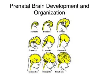
Neural tube defect includes Open and closed neural tube defect. The most common example of neural tube defects includes anencephaly, spina bifida, craniorachischisis, encephalocele, iniencephaly.

E N D
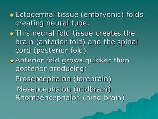
CASE REPORT A rare case of a fetal neural tube defect: Iniencephaly Iniencephaly is characterized by severe retro flexion of the head with the absence of neck due to spinal vertebrae deformities, it is considered as uncommon anomaly. Prevalence: 0.1-10:10,000, M:F. Etiology: Unknown, but genetic, environmental factors are implicated. Pathogenesis: Unknown. Recurrence risk: 1-4%. Associated anomalies: Anencephaly, encephalocele, cyclopedia, lack of lower jaw bone, cleft palate, arthrogryposis, clubfeet, holoprosencephaly, spina bifida, lung hypoplasia, omphalocele, gastroschisis, cardiovascular disorders, Congenital diaphragmatic hernias, gastrointestinal atresia, single umbilical artery and renal abnormalities. Conclusion: Iniencephaly is a lethal congenital neural tube malformation. It is characterized by occipital bone defect, fixed retroflexion of the fetal head and severe lordosis of the cervicothoracic spine. Differential diagnosis: Include anencephaly with cervical spinal retro flexion and Klippel-Fiel syndrome. KEYWORDS: neural tube defectiniencephalyspina pifidaanencephaly of the two eye cavities into one), lack of lower jaw bone, cleft palate, arthrogryposis, clubfeet, holoprosencephaly, spina bifida, low set ears, pulmonary hypoplasia, omphalocele, gastroschisis, cardiovascular disorders, diaphragmatic hernias, gastrointestinal atresia, single umbilical artery and renal abnormalities [1]. Introduction Neural tube defect includes Open and closed neural tube defect. The most common example of neural tube defects includes anencephaly, spina bifida, craniorachischisis, encephalocele, iniencephaly. Sherif Elsirgany*1, Sameh Salama1, Mahmoud Al Alfy1 & Mona Aboulghar2 1Reproductive Health Research Department, National Research Centre, Cairo, Egypt Anencephaly is a defect in the development of the central nervous system in which the Brain tissue and cranial vault are grossly deformed. Case Report With a history of a 21 years old female, married for one year. She married her cousin. She miscarried her first pregnancy 5 months ago where she was diagnosed as blighted ovum at 7 weeks. She was scheduled for first trimester ultrasound scan in her next pregnancy. She attended her first trimester ultrasound scan appointment in the fetal medicine unit, Obstetrics and Gynecology hospital, Cairo University in her 11th weeks of pregnancy by first day of her period. Ultrasonography was done using Voluson 730 pro machine G.E with 3.5 MHz linear - transducer. The scan revealed: 2Obstetrics & Gynecology Department, Cairo University, Egypt Craniorachischisis is congenital fissure of the skull and vertebral column [1]. This defect results when the neural tube fails to close during the third to fourth weeks of gestation, leading to fetal loss & stillbirth. *Author for correspondence: sherifelsirgany@yahoo.com Iniencephaly is a rare and lethal congenital malformation of the neural tube characterized by occipital bone defect, cervical dysraphism, fixed retroflexion of the fetal head and severe lordosis of the cervicothoracic spine [2]. There are two types of iniencephaly [3]. Iniencephaly apertus, which is associated with encephalocele is considered the more severe type. The other group is not associated with the encephalocele (iniencephaly Iniencephaly may be anencephaly, encephalocele (cranial brain tissue extrudes from the skull), cyclopedia (fusion 1) Single living intrauterine fetus with Crown Rump Length (CRL)=44 mm. clauses). with 2) Short rotated spine, fixed retroflexed head, occipital encephalocele, arthrogryposis and omphalocele (FIGURE 1). accompanied 17 Imaging Med.(2018) 10(1) ISSN 1755-5191
CASE REPORT Elsirgany, Salama, Al Alfy, Aboulghar Figure 1. Ultrasonography examination of fetal neural tube defect: Iniencephaly. These findings were constant after repeating the scan after an hour. The head was larger than the trunk and abdomen, disproportionately. The head was in fixed hyperextension and no neck region was visualized. After confirmation of diagnosis, the condition was explained to the patient with its high mortality rate. Termination of pregnancy has been suggested and the procedure has been explained to the patient. The patient accepted and consented for termination of her pregnancy which has been two days later. Medical termination of pregnancy was done by using vaginal misoprostol 200 µg. Postmortem Chromosomal examination has been offered to the patient and she agreed and consented. The chromosomal study was done and revealed normal female karyotype (46,XX) (FIGURE 1). of the neural tube is normally completed by 28 days after conception [5]. Iniencephaly is a rare neural tube defect (NTD). Incidence ranges from 0.1 to 10 in 10,000. It is more common in females (about 90%), once a mother has given birth to a child with iniencephaly, risk of recurrence increases to 1-5% [6]. Until now, the exact etiology and pathogenesis is not known, both genetic and environmental causes may have a role. Chromosomal abnormalities including trisomy 18, trisomy 13 and monosomy X have been detected with this anomaly [7]. Environmental factors such as low socioeconomic conditions, low parity, and deficiency of folic acid supplementation, obesity and drugs including sulphonamide, tetracycline, antihistamines, clomiphen citrate and antitumor agents are shown to have increased risk. It was noticed that women with hyperhomocysteinemia associated with increase the risk of neural tube defect in her child birth. Folic acid decreases the raised homocystein levels and decreases the risk of neural tube defect (NTDs). Discussion The terminology of Iniencephaly derived from Greek word inion which refers to back of the neck and encephalos refer to brain. The fusion of the posterior most part of occipital bone with the back leading to the absence of the neck and retroflexion of head [4]. The diagnosis of iniencephaly antenatally by During embryological life, the development 18 Imaging Med.(2018) 10(1)
CASE REPORT A rare case of a fetal neural tube defect: Iniencephaly ultrasonography/Magnetic Resonance Imaging (MRI) or Computed Tomography (CT). The fetus will show typical star gazing appearance on USG and detailed CNS and spine abnormalities may be known by Magnetic Resonance Imaging (MRI) or Computed Tomography (CT) [8]. (KFS) and cervical meningomyelocele [9]. Anencephaly shows a total or partial absence of neurocranium and retroflexed head is not covered with skin. However, in iniencephaly the retroflexed head is completely covered with skin. Cervical vertebrae are abnormal in iniencephaly and they are almost normal in anencephaly. In the present case the retroflexed head was completely covered with skin and there was encephalocele. Klippel-Fiel syndrome is characterized by the congenital fusion of any 2 of the 7 cervical vertebrae. The keys for diagnosis a case of iniencephaly by ultrasound includes: 1. Irregular fusion of malformed vertebrae. 2. Incomplete closure of vertebral arches and bodies. Prognosis Iniencephaly apertus is always fatal in the neonatal period [10] and our case was diagnosed as iniencephaly apertus and the termination of pregnancy was suggested and the procedure was explained to her. Four cases with a mild iniencephalus clausus have been reported with long-term survival, although in these cases, the deformity was minimal and they should probably have been classified as Klippel-Feil syndrome [11]. 3. Retroflexion of the cervical spine. Upward turned face with chin continuous with chest because of the absence of neck [9]. Our case had all these features and hence was diagnosed as iniencephaly. Differential diagnosis Iniencephaly apertus should be differentiated from anencephaly with spine. Also, iniencephaly clausus should be differentiated from Klippel-Fiel syndrome retroflexion of Sonographic and MR correlation: A case report. Korean. J. Radiol. 8, 351-355 (2004). 8. Gadodia A, Gupta P, Sharma R et al. Antenatal sonography and MRI of iniencephaly apertus and clausus. Fetal. Diagn. Ther. 27, 178-180 (2010). REFERENCES 1. Baker PN. Prenatal diagnosis. In: Obstetrics by 10 teachers. Book. Power. Publishers. Hodder Arnold. London. 18, 107-108 (2006). 5. Botto LD, Moore CA, Khoury MJ. Neuraltube defects. N. Engl. J. Med. 341, 1509-1519 (1999). 9. Chen CP. Prenatal diagnosis of iniencephaly. Taiwan. J. Obstet. Gynecol. 46, 199-207 (2007). 2. Balci S, Aypar E, Altinok G et al. Prenatal diagnosis in three cases of iniencephaly with unusual postmortem findings. Prenat. Diagn. 21, 558-562 (2001). 6. Pungavkar iniencephaly: Sonographic and MR correlation: A case report. Korean. J. Radiol. 8, 351-355 (2007). SA. Antenatal diagnosis of 10. Nishimura H, Okamoto N. Iniencephaly - Handbook of Clinical Neurology. New. York. North Holland. Biochemical. Press. 30, 257- 268 (1977). 3. Moore CA. Neural tube defects from origin to treatment. Cerebrospinal. Fluid. Res. 3, 6 (2006). 7. Semi T, Mehmet U, Oya P et al. Iniencephaly: Prenatal diagnosis with postmortem findings. J. Obstet. Gybocol. 33, 5669 (2007). 11. Katz VL, Aylsworth AS, Albright SG: Iniencephaly is not uniformly fatal. Prenat. Diagn. 9, 595-599 (1989). 4. Pungavkar SA, Sainani NI, Karnik AS et al. Antenatal diagnosis of iniencephaly: 19 Imaging Med.(2018) 10(1)