Figure 2.1. CSE separation of eight molecular weight standards.
38. Figure 2.1. CSE separation of eight molecular weight standards. Capillary : coated 40-cm x 50- m ID. Injection : -125-V/cm for 3-s; E = -500-V/cm across the capillary. Sieving matrix : 0.1-M CHES + 0.1-M Tris and 6- % pullulan with 3.5-mM SDS at pH 8.7.
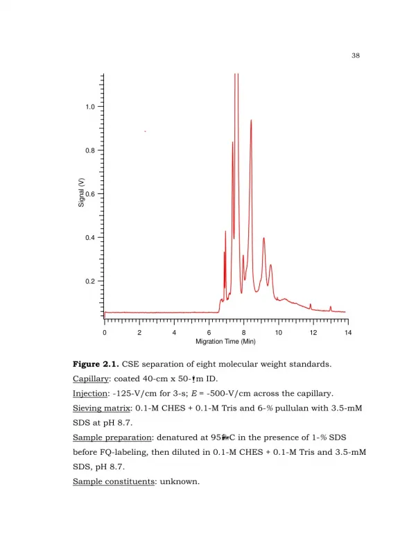

Figure 2.1. CSE separation of eight molecular weight standards.
E N D
Presentation Transcript
38 Figure 2.1. CSE separation of eight molecular weight standards. Capillary: coated 40-cm x 50-m ID. Injection: -125-V/cm for 3-s; E = -500-V/cm across the capillary. Sieving matrix: 0.1-M CHES + 0.1-M Tris and 6-% pullulan with 3.5-mM SDS at pH 8.7. Sample preparation: denatured at 95C in the presence of 1-% SDS before FQ-labeling, then diluted in 0.1-M CHES + 0.1-M Tris and 3.5-mM SDS, pH 8.7. Sample constituents: unknown.
38 -Lactoglobulin MW = 18,281-Da Figure 2.2. CSE separation of -lactoglobulin in pullulan. Capillary: coated 40-cm x 50-m ID. Injection: -50-V/cm for 3-s; E = -300-V/cm across the capillary. Sieving matrix: 0.1-M CHES + 0.1-M Tris and 6-% pullulan with 3.5-mM SDS at pH 8.7. Sample preparation: -lactoglobulin was denatured at 95C in the presence of 1-% SDS before FQ-labeling, then diluted in 0.1-M CHES + 0.1-M Tris and 3.5-mM SDS, pH 8.7 to a concentration of 50-nM.
38 -lactoglobulin MW = 18,281-Da Figure 2.3. CSE separation of -lactoglobulin in pullulan. Capillary: coated 40-cm x 50-m ID. Injection: -50-V/cm for 3-s; E = -350-V/cm across the capillary. Sieving matrix: 0.1-M CHES + 0.1-M Tris and 6-% pullulan with 3.5-mM SDS at pH 8.7. Sample preparation: -lactoglobulin was denatured at 95C in the presence of 1-% SDS before FQ-labeling, then diluted in 0.1-M CHES + 0.1-M Tris and 3.5-mM SDS, pH 8.7 to a concentration of 50-nM.
38 BSA MW = 66,432-Da Figure 2.4. CSE separation of BSA in pullulan. Capillary: coated 40-cm x 50-m ID. Injection: -50-V/cm for 5-s; E = -350-V/cm across the capillary. Sieving matrix: 0.1-M CHES + 0.1-M Tris and 6-% pullulan with 3.5-mM SDS at pH 8.7. Sample preparation: BSA was denatured at 95C in the presence of 1-% SDS before FQ-labeling, then diluted in 0.1-M CHES + 0.1-M Tris and 3.5-mM SDS, pH 8.7 to a concentration of 100-nM.
38 Ovalbumin MW = 42,750-Da Figure 2.5. CSE separation of ovalbumin in pullulan. Capillary: coated 40-cm x 50-m ID. Injection: -50-V/cm for 5-s; E = -350-V/cm across the capillary. Sieving matrix: 0.1-M CHES + 0.1-M Tris and 6-% pullulan with 3.5-mM SDS at pH 8.7. Sample preparation: ovalbumin was denatured at 95C in the presence of 1-% SDS before FQ-labeling, then diluted in 0.1-M CHES + 0.1-M Tris and 3.5-mM SDS, pH 8.7 to a concentration of 60-nM.
38 Carbonic anhydrase MW = 28,980-Da Figure 2.6. CSE separation of carbonic anhydrase in pullulan. Capillary: coated 40-cm x 50-m ID. Injection: -50-V/cm for 5-s; E = -350-V/cm across the capillary. Sieving matrix: 0.1-M CHES + 0.1-M Tris and 6-% pullulan with 3.5-mM SDS at pH 8.7. Sample preparation: carbonic anhydrase was denatured at 95C in the presence of 1-% SDS before FQ-labeling, then diluted in 0.1-M CHES + 0.1-M Tris and 3.5-mM SDS, pH 8.7 to a concentration of 40-nM.
38 -Lactalbumin MW = 14,186-Da Figure 2.7. CSE separation of -lactalbumin in pullulan. Capillary: coated 40-cm x 50-m ID. Injection: -50-V/cm for 5-s; E = -350-V/cm across the capillary. Sieving matrix: 0.1-M CHES + 0.1-M Tris and 6-% pullulan with 3.5-mM SDS at pH 8.7. Sample preparation: carbonic anhydrase was denatured at 95C in the presence of 1-% SDS before FQ-labeling, then diluted in 0.1-M CHES + 0.1-M Tris and 3.5-mM SDS, pH 8.7 to a concentration of 100-nM.
38 2 4 1 3 5 Figure 2.8. CSE separation of five molecular weight standards. Capillary: coated 40-cm x 50-m ID. Injection: -50-V/cm for 3-s; E = -350-V/cm across the capillary. Sieving matrix: 0.1-M CHES + 0.1-M Tris and 6-% pullulan with 3.5-mM SDS at pH 8.7. Sample preparation: denatured at 95C in the presence of 1-% SDS before FQ-labeling, then diluted in 0.1-M CHES + 0.1-M Tris and 3.5-mM SDS, pH 8.7 to a concentration of 20-nM. Peaks: 1 - -lactoglobulin; 2 - carbonic anhydrase; 3 - ovalbumin; 4 - BSA; 5 - phosphorylase b.
38 38 Figure 2.9. Relationship between the migration time and molecular weight of SDS-denatured protein standards separated in CSE with different sieving matrix concentrations.
38 - Galactosidase MW = 116,351-Da Figure 2.10. CSE separation of -galactosidase in pullulan. Capillary: coated 40-cm x 50-m ID. Injection: -250-V/cm for 5-s; E = -350-V/cm across the capillary. Sieving matrix: 0.1-M CHES + 0.1-M Tris and 6-% pullulan with 3.5-mM SDS at pH 8.7. Sample preparation: -galactosidase was denatured at 95C in the presence of 1-% SDS before FQ-labeling, then diluted in 0.1-M CHES + 0.1-M Tris and 3.5-mM SDS, pH 8.7 to a concentration of 200-nM.
38 - Galactosidase MW = 116,351-Da Figure 2.11. CSE separation of -galactosidase in PEO. Capillary: coated 40-cm x 50-m ID. Injection: -150-V/cm for 5-s; E = -350-V/cm across the capillary. Sieving matrix: 0.1-M CHES + 0.1-M Tris and 2-% PEO(100-kDa) with 3.5-mM SDS at pH 8.7. Sample preparation: -galactosidase was denatured at 95C in the presence of 1-% SDS before FQ-labeling, then diluted in 0.1-M CHES + 0.1-M Tris and 3.5-mM SDS, pH 8.7 to a concentration of 200-nM.
38 5-% dextran 10-% dextran Figure 2.12. CSE separation of -galactosidase in dextran. Capillary: coated 40-cm x 50-m ID. Injection: -150-V/cm for 5-s; E = -350-V/cm across the capillary. Sieving matrix: 60-mM AMPD + 60-mM CACO and dextran(513-kDa) with 3.5-mM SDS at pH 8.7. Sample preparation: -galactosidase was denatured at 95C in the presence of 1-% SDS before FQ-labeling, then diluted in 0.1-M CHES + 0.1-M Tris and 3.5-mM SDS, pH 8.7 to a concentration of 200-nM.
38 5-% dextran N = 4,000-plates N = 1,500-plates 10-% dextran Figure 2.13. CSE separation of -galactosidase in dextran. Capillary: coated 40-cm x 50-m ID. Injection: -150-V/cm for 5-s; E = -350-V/cm across the capillary. Sieving matrix: 0.1-M CHES + 0.1-M Tris and dextran(513-kDa) with 3.5-mM SDS at pH 8.7. Sample preparation: -galactosidase was denatured at 95C in the presence of 1-% SDS before FQ-labeling, then diluted in 0.1-M CHES + 0.1-M Tris and 3.5-mM SDS, pH 8.7 to a concentration of 200-nM.
38 - Galactosidase MW = 116,351-Da Figure 2.14. CSE separation of -galactosidase in PEO. Capillary: coated 40-cm x 50-m ID. Injection: -150-V/cm for 5-s; E = -350-V/cm across the capillary. Sieving matrix: 60-mM AMPD + 60-mM CACO and 2-% PEO(100-kDa) with 3.5-mM SDS at pH 8.8. Sample preparation: -galactosidase was denatured at 95C in the presence of 1-% SDS before FQ-labeling, then diluted in 0.1-M CHES + 0.1-M Tris and 3.5-mM SDS, pH 8.7 to a concentration of 200-nM.
38 2 1 Figure 2.15. CSE separation of 14.2-kDa and 18.4-kDa molecular weight standards in pullulan. Capillary: coated 40-cm x 50-m ID. Injection: -150-V/cm for 5-s; E = -350-V/cm across the capillary. Sieving matrix: 0.1-M CHES + 0.1-M Tris and 6-% pullulan with 3.5-mM SDS at pH 8.7. Sample preparation: denatured at 95C in the presence of 1-% SDS before FQ-labeling, then diluted in 0.1-M CHES + 0.1-M Tris and 3.5-mM SDS, pH 8.7 to a concentration of 50-nM. Peaks: 1 - -lactalbumin; 2 - -lactoglobulin.
38 2 1 RS = 0.502 RS = 0.760 1 2 5-% dextran 10-% dextran Figure 2.16. CSE separation of 14.2-kDa and 18.4-kDa molecular weight standards in pullulan. Capillary: coated 40-cm x 50-m ID. Injection: -150-V/cm for 5-s; E = -350-V/cm across the capillary. Sieving matrix: 0.1-M CHES + 0.1-M Tris and dextran(513-kDa) with 3.5-mM SDS at pH 8.7. Sample preparation: denatured at 95C in the presence of 1-% SDS before FQ-labeling, then diluted in 0.1-M CHES + 0.1-M Tris and 3.5-mM SDS, pH 8.7 to a concentration of 50-nM. Peaks: 1 - -lactalbumin; 2 - -lactoglobulin.
38 1 2 1 2 5-% dextran 10-% dextran Figure 2.17. CSE separation of 14.2-kDa and 18.4-kDa molecular weight standards in pullulan. Capillary: coated 40-cm x 50-m ID. Injection: -150-V/cm for 5-s; E = -350-V/cm across the capillary. Sieving matrix: 60-mM AMPD + 60-mM CACO and dextran(513-kDa) with 3.5-mM SDS at pH 8.8. Sample preparation: denatured at 95C in the presence of 1-% SDS before FQ-labeling, then diluted in 0.1-M CHES + 0.1-M Tris and 3.5-mM SDS, pH 8.7 to a concentration of 50-nM. Peaks: 1 - -lactalbumin; 2 - -lactoglobulin.
38 1 2 3-% PEO 1.5-% PEO Figure 2.18. CSE separation of 14.2-kDa and 18.4-kDa molecular weight standards in pullulan. Capillary: coated 40-cm x 50-m ID. Injection: -150-V/cm for 5-s; E = -350-V/cm across the capillary. Sieving matrix: 0.1-M CHES + 0.1-M Tris and PEO(100-kDa) with 3.5-mM SDS at pH 8.7. Sample preparation: denatured at 95C in the presence of 1-% SDS before FQ-labeling, then diluted in 0.1-M CHES + 0.1-M Tris and 3.5-mM SDS, pH 8.7 to a concentration of 50-nM. Peaks: 1 - -lactalbumin; 2 - -lactoglobulin.
38 1 2 3 Figure 2.19. CSE separation of three molecular weight standards. Capillary: coated 40-cm x 50-m ID. Injection: -150-V/cm for 5-s; E = -350-V/cm across the capillary. Sieving matrix: 0.1-M CHES + 0.1-M Tris and 10-% dextran(513-kDa) with 3.5-mM SDS at pH 8.7. Sample preparation: denatured at 95C in the presence of 1-% SDS before FQ-labeling, then diluted in 0.1-M CHES + 0.1-M Tris and 3.5-mM SDS, pH 8.7. Peaks: 1 - -lactalbumin (50-nM); 2 - -lactoglobulin (10-nM); 3 - carbonic anhydrase (10-nM).
38 1 3 2 Figure 2.20. CSE separation of three molecular weight standards. Capillary: coated 40-cm x 50-m ID. Injection: -150-V/cm for 5-s; E = -350-V/cm across the capillary. Sieving matrix: 0.1-M CHES + 0.1-M Tris and 6-% pullulan with 3.5-mM SDS at pH 8.7. Sample preparation: denatured at 95C in the presence of 1-% SDS before FQ-labeling, then diluted in 0.1-M CHES + 0.1-M Tris and 3.5-mM SDS, pH 8.7. Peaks: 1 - -lactalbumin (50-nM); 2 - -lactoglobulin (10-nM); 3 - carbonic anhydrase (10-nM).
38 2-% PEO 3-% PEO Figure 2.21. CSE separation of three molecular weight standards. Capillary: coated 40-cm x 50-m ID. Injection: -150-V/cm for 5-s; E = -350-V/cm across the capillary. Sieving matrix: 0.1-M CHES + 0.1-M Tris and PEO(100-kDa) with 3.5-mM SDS at pH 8.7. Sample preparation: denatured at 95C in the presence of 1-% SDS before FQ-labeling, then diluted in 0.1-M CHES + 0.1-M Tris and 3.5-mM SDS, pH 8.7. Peaks: 1 - -lactalbumin (50-nM); 2 - -lactoglobulin (10-nM); 3 - carbonic anhydrase (10-nM).
38 1 2 Figure 2.22. CSE separation of two molecular weight standards. Capillary: coated 40-cm x 50-m ID. Injection: -25-V/cm for 5-s; E = -350-V/cm across the capillary. Sieving matrix: 0.1-M CHES + 0.1-M Tris and 6-% pullulan with 3.5-mM SDS at pH 8.7. Sample preparation: denatured at 95C in the presence of 1-% SDS before FQ-labeling, then diluted in 0.1-M CHES + 0.1-M Tris and 3.5-mM SDS, pH 8.7. Peaks: 1 - -lactalbumin (40-nM); 2 - ovalbumin (20-nM).
38 1 3 4 2 Figure 2.23. CSE separation of four molecular weight standards. Capillary: coated 40-cm x 50-m ID. Injection: -25-V/cm for 5-s; E = -350-V/cm across the capillary. Sieving matrix: 0.1-M CHES + 0.1-M Tris and 6-% pullulan with 3.5-mM SDS at pH 8.7. Sample preparation: denatured at 95C in the presence of 1-% SDS before FQ-labeling, then diluted in 0.1-M CHES + 0.1-M Tris and 3.5-mM SDS, pH 8.7 at a concentration of 40-nM. Peaks: 1 - -lactalbumin; 2 - -lactoglobulin; 3 - carbonic anhydrase; 4 - ovalbumin.
38 3-% PEO 2-% PEO Figure 2.24. CSE separation of four molecular weight standards. Capillary: coated 40-cm x 50-m ID. Injection: -25-V/cm for 5-s; E = -350-V/cm across the capillary. Sieving matrix: 60-mM AMPD + 60-mM CACO and PEO(100-kDa) with 3.5-mM SDS at pH 8.8. Sample preparation: denatured at 95C in the presence of 1-% SDS before FQ-labeling, then diluted in 0.1-M CHES + 0.1-M Tris and 3.5-mM SDS, pH 8.7 at a concentration of 40-nM. Sample: -lactalbumin, -lactoglobulin, carbonic anhydrase and ovalbumin; peaks were not identified.
38 1 3 4 2 A B Figure 2.25. CSE separation of four molecular weight standards in 7.5-% dextran(513-kDa). Capillary: coated 40-cm x 50-m ID. Injection: -50-V/cm for 5-s; E = -350-V/cm across the capillary. Sieving matrix: A - 0.1-M CHES + 0.1-M Tris and 3.5-mM SDS at pH 8.7; B - 60-mM AMPD + 60-mM CACO and 3.5-mM SDS at pH 8.8. Sample preparation: denatured at 95C in the presence of 1-% SDS before FQ-labeling, then diluted in 0.1-M CHES + 0.1-M Tris and 3.5-mM SDS, pH 8.7 at a concentration of 40-nM. Peaks: 1 - -lactalbumin; 2 - -lactoglobulin; 3 - carbonic anhydrase; 4 - ovalbumin.
38 1 2 3 4 B A Figure 2.26. CSE separation of four molecular weight standards in 7.5-% dextran(513-kDa). Capillary: coated 40-cm x 50-m ID. Injection: -50-V/cm for 5-s; E = -350-V/cm across the capillary. Sieving matrix: A - 0.1-M CHES + 0.1-M Tris and 3.5-mM SDS at pH 8.7; B - 60-mM AMPD + 60-mM CACO and 3.5-mM SDS at pH 8.8. Sample preparation: denatured at 95C in the presence of 1-% SDS before FQ-labeling, then diluted in 0.1-M CHES + 0.1-M Tris and 3.5-mM SDS, pH 8.7 at a concentration of 40-nM. Peaks: 1 - -lactalbumin; 2 - BSA; 3 - phosphorylase b; 4 - -galactosidase.
38 4 6 2 3 1 5 7 Figure 2.27. CSE separation of seven molecular weight standards. Capillary: coated 40-cm x 50-m ID. Injection: -25-V/cm for 5-s; E = -350-V/cm across the capillary. Sieving matrix: 0.1-M CHES + 0.1-M Tris and 6-% pullulan with 3.5-mM SDS at pH 8.7. Sample preparation: denatured at 95C in the presence of 1-% SDS before FQ-labeling, then diluted in 0.1-M CHES + 0.1-M Tris and 3.5-mM SDS, pH 8.7 at a concentration of 40-nM. Peaks: 1 - -lactalbumin; 2 - -lactoglobulin; 3 - carbonic anhydrase; 4 - ovalbumin; 5 - BSA; 6 - phosphorylase b; 7 - -galactosidase.
38 1) 22,000-plates 2) 17,000-plates 3) 53,000-plates 4) 6,000-plates 5) 7,700-plates 6) 10,300-plates 7) 10,400-plates RS = 0.899 RS = 0.613 RS = 0.748 Figure 2.28. CSE separation of seven molecular weight standards. Capillary: coated 40-cm x 50-m ID. Injection: -25-V/cm for 5-s; E = -350-V/cm across the capillary. Sieving matrix: 60-mM AMPD + 60-mM CACO and 5-% dextran (513-kDa) with 3.5-mM SDS at pH 8.8. Sample preparation: denatured at 95C in the presence of 1-% SDS before FQ-labeling, then diluted in 0.1-M CHES + 0.1-M Tris and 3.5-mM SDS, pH 8.7 at a concentration of 40-nM.
38 3 1 5 2 4 7 6 5-% 6-% 7-% 8-% Figure 2.29. CSE separation of seven molecular weight standards. Capillary: coated 40-cm x 50-m ID. Injection: -25-V/cm for 5-s; E = -350-V/cm across the capillary. Sieving matrix: 60-mM AMPD + 60-mM CACO and dextran(513-kDa) with 3.5-mM SDS at pH 8.8. Sample preparation: denatured at 95C in the presence of 1-% SDS before FQ-labeling, then diluted in 0.1-M CHES + 0.1-M Tris and 3.5-mM SDS, pH 8.7 at a concentration of 40-nM. Peaks: 1 - -lactalbumin; 2 - -lactoglobulin; 3 - carbonic anhydrase; 4 - ovalbumin; 5 - BSA; 6 - phosphorylase b; 7 - -galactosidase.
38 3 6 1 7 2 5 4 5-% 4-% 3-% Figure 2.30. CSE separation of seven molecular weight standards. Capillary: coated 40-cm x 50-m ID. Injection: -50-V/cm for 5-s; E = -350-V/cm across the capillary. Sieving matrix: 60-mM AMPD + 60-mM CACO and dextran(2-MDa) with 3.5-mM SDS at pH 8.8. Sample preparation: denatured at 95C in the presence of 1-% SDS before FQ-labeling, then diluted in 0.1-M CHES + 0.1-M Tris and 3.5-mM SDS, pH 8.7 at a concentration of 40-nM. Peaks: 1 - -lactalbumin; 2 - -lactoglobulin; 3 - carbonic anhydrase; 4 - ovalbumin; 5 - BSA; 6 - phosphorylase b; 7 - -galactosidase.
38 1 2 4 3 6 5 7 Figure 2.31. CSE separation of seven molecular weight standards. Capillary: coated 40-cm x 50-m ID. Injection: -50-V/cm for 5-s; E = -500-V/cm across the capillary. Sieving matrix: 0.1-M CHES + 0.1-M Tris and 7-% dextran(513-kDa) with 3.5-mM SDS at pH 8.7. Sample preparation: denatured at 95C in the presence of 1-% SDS before FQ-labeling, then diluted in 0.1-M CHES + 0.1-M Tris and 3.5-mM SDS, pH 8.7 at a concentration of 40-nM. Peaks: 1 - -lactalbumin; 2 - -lactoglobulin; 3 - carbonic anhydrase; 4 - ovalbumin; 5 - BSA; 6 - phosphorylase b; 7 - -galactosidase.
38 2 6 4 3 1 5 7 Figure 2.32. CSE separation of seven molecular weight standards in dextran-pullulan sieving mixture. Capillary: coated 40-cm x 50-m ID. Injection: -25-V/cm for 5-s; E = -350-V/cm across the capillary. Sieving matrix: 0.1-M CHES + 0.1-M Tris and 2.5-% dextran(513-kDa) + 2.5-% pullulan with 3.5-mM SDS at pH 8.7. Sample preparation: denatured at 95C in the presence of 1-% SDS before FQ-labeling, then diluted in 0.1-M CHES + 0.1-M Tris and 3.5-mM SDS, pH 8.7 at a concentration of 40-nM. Peaks: 1 - -lactalbumin; 2 - -lactoglobulin; 3 - carbonic anhydrase; 4 - ovalbumin; 5 - BSA; 6 - phosphorylase b; 7 - -galactosidase.
38 3 6 1 2 7 5 4 Figure 2.33. CSE separation of seven molecular weight standards in dextran-pullulan sieving mixture. Capillary: coated 40-cm x 50-m ID. Injection: -25-V/cm for 5-s; E = -450-V/cm across the capillary. Sieving matrix: 60-mM AMPD + 60-mM CACO and 2-% dextran(513-kDa) + 2-% dextran(2-MDa) + 2-% pullulan with 3.5-mM SDS at pH 8.8. Sample preparation: denatured at 95C in the presence of 1-% SDS before FQ-labeling, then diluted in 0.1-M CHES + 0.1-M Tris and 3.5-mM SDS, pH 8.7 at a concentration of 40-nM. Peaks: 1 - -lactalbumin; 2 - -lactoglobulin; 3 - carbonic anhydrase; 4 - ovalbumin; 5 - BSA; 6 - phosphorylase b; 7 - -galactosidase.
38 2 1 6 3 5 7 4 5-% 4-% 3-% Figure 2.34. CSE separation of seven molecular weight standards. Capillary: coated 40-cm x 50-m ID. Injection: -25-V/cm for 5-s; E = -350-V/cm across the capillary. Sieving matrix: 0.1-M CHES + 0.1-M Tris and dextran(2-MDa) with 3.5-mM SDS at pH 8.7. Sample preparation: denatured at 95C in the presence of 1-% SDS before FQ-labeling, then diluted in 0.1-M CHES + 0.1-M Tris and 3.5-mM SDS, pH 8.7 at a concentration of 40-nM. Peaks: 1 - -lactalbumin; 2 - -lactoglobulin; 3 - carbonic anhydrase; 4 - ovalbumin; 5 - BSA; 6 - phosphorylase b; 7 - -galactosidase.
38 2 6 3 5 1 4 7 A B C Figure 2.35. CSE separation of seven molecular weight standards in dextran-pullulan sieving mixture. Capillary: coated 40-cm x 50-m ID. Injection: -25-V/cm for 5-s; E = -450-V/cm across the capillary. Sieving matrix: A - 2-% dextran(513-kDa) + 2-% pullulan; B - 2-% dextran(2-MDa) + 2-% pullulan; C - 2-% dextran(513-kDa) + 2-% dextran(2-MDa). Sample preparation: denatured at 95C in the presence of 1-% SDS before FQ-labeling, then diluted in 0.1-M CHES + 0.1-M Tris and 3.5-mM SDS, pH 8.7 at a concentration of 40-nM. Peaks: 1 - -lactalbumin; 2 - -lactoglobulin; 3 - carbonic anhydrase; 4 - ovalbumin; 5 - BSA; 6 - phosphorylase b; 7 - -galactosidase.
38 2 1 6 3 4 5 7 Figure 2.36. CSE separation of seven molecular weight standards in dextran-pullulan sieving mixture. Capillary: coated 40-cm x 50-m ID. Injection: -25-V/cm for 5-s; E = -450-V/cm across the capillary. Sieving matrix: 0.1-M CHES + 0.1-M Tris and 2-% dextran(513-kDa) + 2-% dextran(2-MDa) + 2-% pullulan with 3.5-mM SDS at pH 8.7. Sample preparation: denatured at 95C in the presence of 1-% SDS before FQ-labeling, then diluted in 0.1-M CHES + 0.1-M Tris and 3.5-mM SDS, pH 8.7 at a concentration of 40-nM. Peaks: 1 - -lactalbumin; 2 - -lactoglobulin; 3 - carbonic anhydrase; 4 - ovalbumin; 5 - BSA; 6 - phosphorylase b; 7 - -galactosidase.
38 Figure 2.37. CSE separation of five molecular weight standards. Capillary: coated 40-cm x 50-m ID. Injection: -100-V/cm for 5-s; E = -500-V/cm across the capillary. Sieving matrix: 25-mM CHES + 25-mM Tris and 5-% dextran(513-kDa) with 3.5-mM SDS at pH 8.7. Sample preparation: denatured at 95C in the presence of 1-% SDS before FQ-labeling, then diluted in 0.1-M CHES + 0.1-M Tris and 3.5-mM SDS, pH 8.7 at a concentration of 40-nM. Sample: carbonic anhydrase, ovalbumin, BSA, phosphorylase b, -galactosidase; peaks were not identified.
38 1 3 2 4 5 Figure 2.38. CSE separation of five molecular weight standards. Capillary: coated 40-cm x 50-m ID. Injection: -125-V/cm for 2-s; E = -375-V/cm across the capillary. Sieving matrix: 0.1-M CHES + 0.1-M Tris and 5-% dextran(513-kDa) with 3.5-mM SDS at pH 8.7. Sample preparation: denatured at 95C in the presence of 1-% SDS before FQ-labeling, then diluted in 0.1-M CHES + 0.1-M Tris and 3.5-mM SDS, pH 8.7 at a concentration of 40-nM. Peaks: 1 - carbonic anhydrase; 2 - ovalbumin; 3 - BSA; 4 - phosphorylase b; 5 - -galactosidase.
38 1 3 2 4 5 X = 100-mM X = 75-mM X = 50-mM X = 25-mM Figure 2.39. CSE separation of five molecular weight standards in various buffer concentrations. Capillary: coated 40-cm x 50-m ID. Injection: -100-V/cm for 2-s; E = -500-V/cm across the capillary. Sieving matrix: X-CHES + X-Tris and 5-% dextran(513-kDa) with 3.5-mM SDS at pH 8.7. Sample preparation: denatured at 95C in the presence of 1-% SDS before FQ-labeling, then diluted in 0.1-M CHES + 0.1-M Tris and 3.5-mM SDS, pH 8.7 at a concentration of 40-nM. Peaks: 1 - carbonic anhydrase; 2 - ovalbumin; 3 - BSA; 4 - phosphorylase b; 5 - -galactosidase.
38 Figure 2.40. CSE separation of ovalbumin (45-kDa) in dextran. Capillary: coated 40-cm x 50-m ID. Injection: -100-V/cm for 2-s; E = -500-V/cm across the capillary. Sieving matrix: 0.1-M CHES + 0.1-M Tris and 5-% dextran(513-kDa) with 3.5-mM SDS at pH 8.7. Sample preparation: ovalbumin was denatured at 95C in the presence of 1-% SDS before FQ-labeling, then diluted in 0.1-M CHES + 0.1-M Tris and 3.5-mM SDS, pH 8.7 at a concentration of 50-nM.
38 Ovalbumin Isolated with ProTip From stock bottle Figure 2.41. CSE separation of ovalbumin from a stock bottle compared to ovalbumin isolated from a ProTip. Capillary: coated 40-cm x 50-m ID. Injection: -25-V/cm for 2-s; E = -375-V/cm across the capillary. Sieving matrix: 0.1-M CHES + 0.1-M Tris and 5-% dextran(513-kDa) with 3.5-mM SDS at pH 8.7. Sample preparation: ovalbumin was heated at 95C in the presence of 1-% SDS prior to FQ-labeling, then diluted in 0.1-M CHES + 0.1-M Tris and 3.5-mM SDS, pH 8.7 at a concentration of 4-M.
38 Lysozyme Figure 2.42. CSE separation of lysozyme in dextran (513-kDa). Capillary: coated 40-cm x 50-m ID. Injection: -75-V/cm for 2-s; E = -375-V/cm across the capillary. Sieving matrix: 0.1-M CHES + 0.1-M Tris and 5-% dextran(513-kDa) with 3.5-mM SDS at pH 8.7. Sample preparation: lysozyme was heated at 95C in the presence of 1-% SDS prior to FQ-labeling, then diluted in 0.1-M CHES + 0.1-M Tris and 3.5-mM SDS, pH 8.7 at a concentration of 700-nM.
38 Myoglobin Figure 2.43. CSE separation of myoglobin in dextran (513-kDa). Capillary: coated 40-cm x 50-m ID. Injection: -75-V/cm for 2-s; E = -375-V/cm across the capillary. Sieving matrix: 0.1-M CHES + 0.1-M Tris and 5-% dextran(513-kDa) with 3.5-mM SDS at pH 8.7. Sample preparation: myoglobin was heated at 95C in the presence of 1-% SDS prior to FQ-labeling, then diluted in 0.1-M CHES + 0.1-M Tris and 3.5-mM SDS, pH 8.7 at a concentration of 150-nM.
38 apo-Myoglobin Figure 2.44. CSE separation of apo-myoglobin in dextran. Capillary: coated 40-cm x 50-m ID. Injection: -75-V/cm for 2-s; E = -375-V/cm across the capillary. Sieving matrix: 0.1-M CHES + 0.1-M Tris and 5-% dextran(513-kDa) with 3.5-mM SDS at pH 8.7. Sample preparation: apo-myoglobin was heated at 95C in the presence of 1-% SDS prior to FQ-labeling, then diluted in 0.1-M CHES + 0.1-M Tris and 3.5-mM SDS, pH 8.7 at a concentration of 180-nM.