Download

1 / 83
0 likes | 1 Views
Fluoroscopy-Free Techniques in Modern Orthopedics<br>

E N D
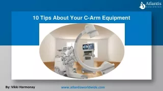
Osteotomies Without C-Arm: Tips and Tricks السلام عليكم ورحمة الله وبركاته Dr. Bahaa Ali Kornah Prof.. Of Orthopedic Al-Azhar University Cairo - Egypt Bahaa Ali Kornah-Al-Azhar Un. Cairo -EGYPT
Dr. Bahaa Ali Kornah • Prof. Of Orthopedic • Al-Azhar University • Cairo Egypt Bahaa Ali Kornah-Al-Azhar Un. Cairo -EGYPT
Osteotomies Without C-Arm: Tips and Tricks Bahaa Ali Kornah-Al-Azhar Un. Cairo -EGYPT
Fluoroscopy-Free Techniques in Modern Orthopedics Bahaa Ali Kornah-Al-Azhar Un. Cairo -EGYPT
Introduction • Importance of fluoroscopy in osteotomies • Limitations: radiation exposure, equipment dependence • When and why to perform without a C-arm • Overview of this presentation Bahaa Ali Kornah-Al-Azhar Un. Cairo -EGYPT
Osteotomy • An osteotomy is a surgical operation whereby a bone is cut to • shorten, • lengthen, or • change its alignment Bahaa Ali Kornah-Al-Azhar Un. Cairo -EGYPT
Indications for Fluoroscopy-Free Osteotomy • Limited access to fluoroscopy (e.g., resource-limited settings) • Preoperative planning with 3D imaging or navigation • Experienced surgeons familiar with anatomy-based techniques • Pediatric or deformity correction cases with clear landmarks Bahaa Ali Kornah-Al-Azhar Un. Cairo -EGYPT
Preoperative Planning • Detailed templating with CT or long-leg X-rays • Identifying deformity type and apex • Marking mechanical and anatomical axes • Planning correction angle and bone cuts Bahaa Ali Kornah-Al-Azhar Un. Cairo -EGYPT
Surgical Anatomy Review • Key bony landmarks: Tibial tuberosity, fibular head, medial malleolus • Distal femur condyles, intercondylar notch • Surface anatomy correlation • Use of intraoperative guides or palpation techniques Bahaa Ali Kornah-Al-Azhar Un. Cairo -EGYPT
Instrumentation & Setup • Long alignment rods • Mechanical axis markers (electromechanical or cord) • Use of osteotomy jigs or cutting guides • Calibration tools for intraoperative assessment Bahaa Ali Kornah-Al-Azhar Un. Cairo -EGYPT
Osteotomy guide system Bahaa Ali Kornah-Al-Azhar Un. Cairo -EGYPT
Double level knee osteotomy using patient-specific cutting guides Bahaa Ali Kornah-Al-Azhar Un. Cairo -EGYPT
Fixation of Osteotomies • Internal fixation • Plates and screws • Intramedullary nails • Staples or lag screws • External fixation • Cast • Monolateral external fixators • Circular (Ilizarov / hexapod) frames • Hybrid fixation • Combination of internal and external fixation Bahaa Ali Kornah-Al-Azhar Un. Cairo -EGYPT
Types ofosteotomies: • A: Openingwedge. • B: Closingwedge. • C:Displacement. • D:Stepcut. • E: Crescentic(dome). • F: Transversederotation. • G: Oblique (singleplane). Bahaa Ali Kornah-Al-Azhar Un. Cairo -EGYPT
TRANSVERSEOSTEOTOMY • Ideal for correcting rotationalone. • Open or Percutaneous • Diaphysisor metaphysis plane of cut is transverse to long axis of bone to avoid frontal or sagittaldeformity. • Simple to performbut it is relatively unstableand is not ideally suited for interfragmentary compression. • Resists axialloadbut weak to torsion orbending loads. • Angularcorrections difficult to control. Bahaa Ali Kornah-Al-Azhar Un. Cairo -EGYPT
CLOSING WEDGE OSTEOTOMY • Technique • Remove a pre-planned bone wedge at maximum deformity • Base of wedge on convex side • Close osteotomy and apply internal fixation • Indication • HTO for unicompartmental knee osteoarthritis • Correction of varus deformity • Advantages • Simple and stable technique • Rapid bone healing • No bone graft required • Disadvantages • Alters soft-tissue balance near joints • Causes limb shortening Bahaa Ali Kornah-Al-Azhar Un. Cairo -EGYPT
OPENINGWEDGEOSTEOTOMY:- • Performed using a single transverse osteotomy • The osteotomy is opened on the concave side and filled with bone graft • Base of the wedge lies on the concave surface • Apex of the wedge lies on the convex surface • Advantages • Allows slight limb lengthening • Enables precise correction • Disadvantages • Requires bone graft • Slower healing compared to closing wedge • Risk of delayed union or non-union • Full weight bearing delayed until graft incorporation and remodeling Bahaa Ali Kornah-Al-Azhar Un. Cairo -EGYPT
OBLIQUE (SINGLE-PLANE)OSTEOTOMY • It can correct all deformities with a singlecut. • broader surface area forhealing. • Compression atsite. • Canlengthen. • No graft . • Creates somerotation. • useful in the metaphysealregion. Bahaa Ali Kornah-Al-Azhar Un. Cairo -EGYPT
CRESCENTIC (DOME)OSTEOTOMY • Used in metaphyseal or epiphyseal cancellous bone • Irregular bone cuts provide good inherent stability • Broad cancellous surface area allows rapid healing • Technique • Dome-shaped osteotomy • One side shallow, the other deep • Deformity corrected by rotational realignment • Advantages • Bone-preserving procedure • Maintains limb length • Indications • Ideal for deformity near joints • Best for deformities in a single plane (especially frontal plane) Bahaa Ali Kornah-Al-Azhar Un. Cairo -EGYPT
DISPLACEMENTOSTEOTOMY • Wagner’s transverse metaphyseal osteotomy corrects major juxtaarticulardeformities • by rotating a periarticular fragment • so one corner impacts into the medullary canal of the adjacent fragment. • Converts bending forces into compression, • Maintains limb length, and restores joint alignment. Bahaa Ali Kornah-Al-Azhar Un. Cairo -EGYPT
STEPCUTOSTEOTOMY:- • A surgical technique involving a stepped (L- or Z-shaped) bone cut • provides enhanced rotational and translational stability • maintain alignment, reduces displacement risk, and often eliminates the need for extensive internal fixation • such as one-stage diaphyseal lengthening. • Rotational and angular corrections are limited. Bahaa Ali Kornah-Al-Azhar Un. Cairo -EGYPT
COMPLICATIONSOFOSTEOTOMYAND DEFORMITYCORRECTION:- • 1. Intraoperative Complications • Neurovascular Injury: Damage to nearby nerves (e.g., peroneal, tibial) or vessels during bone cutting or retraction. • Fracture: Inadvertent fracture due to poor bone quality or excessive force. • Inaccurate Cut/Alignment: Technical error leading to under- or over-correction. • 2. Early Postoperative Complications • Infection (superficial or deep) • Delayed Union / Nonunion: Failure of bone to heal within expected time (often >3–6 months). • Malunion: Healing in incorrect position due to loss of fixation or inadequate correction. • Compartment Syndrome: Especially in tibial osteotomies — requires urgent fasciotomy. • Thromboembolism (DVT/PE): Risk increased with immobility and surgery duration. • Hardware Failure or Irritation: Screws/plates breaking or causing discomfort. • 3. Late Complications • Joint Degeneration: Altered biomechanics may accelerate arthritis in adjacent joints (e.g., knee after tibial osteotomy). • Recurrent Deformity: Due to growth (in pediatric cases), non-compliance, or progressive disease. • Stiffness or Reduced Range of Motion: From scarring, prolonged immobilization, or muscle contractures. • Leg Length Discrepancy: If correction alters mechanical axis significantly without compensation. • Reflex Sympathetic Dystrophy (CRPS): Chronic pain syndrome following trauma/surgery. Bahaa Ali Kornah-Al-Azhar Un. Cairo -EGYPT
Varus Osteotomy – • Purpose & Indications • 🔹 Objective:To elevate and laterally reposition the greater trochanter while shifting the abductor and iliopsoas muscles medially — thereby improving hip joint congruency and reducing abnormal muscle forces acting across the joint. • 🔹 Indications: • Patients with a spherical femoral head and minimal or no acetabular dysplasia (center-edge angle ≥15–20°) • Presence of a valgus neck-shaft angle >135° • Fixed abduction deformity of the hip • 🔹 Contraindications (C/I): • Fixed external rotation >25° • Flexion contracture >70° • 🔹 Mechanical Benefit: • Performing a varus osteotomy with medial displacement of the femoral shaft: • Relaxes the abductors, iliopsoas, and adductors • Reduces joint reactive forces (unloads the hip) • Expands the weight-bearing surface area of the femoral head within the acetabulum Bahaa Ali Kornah-Al-Azhar Un. Cairo -EGYPT
Varus osteotomy increases weight bearing area of femoral head while relaxing all three important muscle groups around hip joint Bahaa Ali Kornah-Al-Azhar Un. Cairo -EGYPT
Three types of wedges cut for varus osteotomy. A, Original technique of Pauwels with proximal osteotomy made transversely at distal end of greater trochanter. This type of osteotomy makes it more difficult to correct rotation and to use right-angled blade plate. B, Original Müller technique of excision of wide wedge based medially with distal osteotomy cut transversely across shaft at just above level of lesser trochanter. C, Later technique of Müller using small half wedge cut medially and transposed laterally. Bahaa Ali Kornah-Al-Azhar Un. Cairo -EGYPT
VARUS OSTEOTOMIES Bahaa Ali Kornah-Al-Azhar Un. Cairo -EGYPT
🔹 Medial Displacement Recommendation: • Most authors suggest a medial displacement of 10–15 mm to: • Keep the ipsilateral knee centered under the femoral head • Preserve the mechanical axis of the leg • 🔹 Consequences of Varus Osteotomy: • Inevitably causes some degree of limb shortening • May result in a Trendelenburg gait that can persist for months post-op • Increases the prominence of the greater trochanter • 🔹 Minimizing Limb Shortening: • Can be reduced by: • Performing a smaller medial osteotomy • Transposing the fragment laterally (instead of purely medially) Bahaa Ali Kornah-Al-Azhar Un. Cairo -EGYPT
CORRECTIVE OSTOTOMIES Whitman closing wedge Brackett ball and socket Gant-opening wedge Osteotomy Osteotomy Osteotomy Bahaa Ali Kornah-Al-Azhar Un. Cairo -EGYPT
Techniques – High Tibial Osteotomy (HTO) • Medial opening wedge vs. lateral closing wedge • Landmark-guided saw cut depth • Use of cables/rods for limb axis assessment • Step-by-step workflow without fluoroscopy Bahaa Ali Kornah-Al-Azhar Un. Cairo -EGYPT
Indications • Unicompartmental Osteoarthritis • Under 60 ys • Good ROM • No Flexion Contracture • Not Overweight • Unloading for Meniscal/Chondral Grafting • Relative for ACL or PLC deficient in Varus Bahaa Ali Kornah-Al-Azhar Un. Cairo -EGYPT
Contra-indications • Bi- or tri-compartmental joint destruction • Lateral OA (clinical results are not predictable) • OCD lesion more than 5mm deep • Flexion contracture exceeding 10 degrees • Overall ROM of less than 90 degrees • Varus deformation of more than 15 degrees • Inadequate bone stock • Instability Bahaa Ali Kornah-Al-Azhar Un. Cairo -EGYPT
Radiographs • Full Length weight bearing AP with patella facing forward • Supine film • 30 degree PA flex • Lateral • Sunrise Bahaa Ali Kornah-Al-Azhar Un. Cairo -EGYPT
Percutaneous tibial osteotomy Bahaa Ali Kornah-Al-Azhar Un. Cairo -EGYPT
Percutaneous tibial osteotomy Bahaa Ali Kornah-Al-Azhar Un. Cairo -EGYPT
Percutaneous tibial osteotomy Bahaa Ali Kornah-Al-Azhar Un. Cairo -EGYPT
Percutaneous tibial osteotomy Bahaa Ali Kornah-Al-Azhar Un. Cairo -EGYPT
Percutaneous tibial osteotomy Bahaa Ali Kornah-Al-Azhar Un. Cairo -EGYPT