Download

1 / 27
270 likes | 579 Views
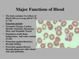
Major components of blood. I. Fluid component - plasma. A. Water - 90%. B. Plasma proteins - 7% a. albumin b. fibrinogen c. alpha, beta, and gamma globulins - gamma globulins are IgG antibodies (immunoglobulins). C. Inorganic salts - 0.9%.

E N D
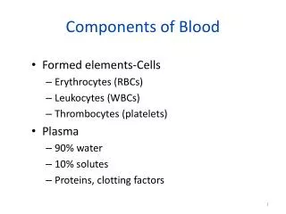
Major components of blood I. Fluid component - plasma. A. Water - 90% B. Plasma proteins - 7% a.albumin b.fibrinogen c.alpha, beta, and gamma globulins - gamma globulins are IgG antibodies (immunoglobulins) C.Inorganic salts - 0.9% D.Other organic molecules - 2.1% (vitamins, amino acids, lipids, hormones, etc.)
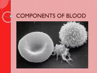
Major components of blood 1. no nucleus in mature erythrocyte 2. biconcave disk shape 3. mature cell is essentially a sac of hemoglobulin 4. 7-8 μm in diameter 5. most common type of blood cell 6. reticulocytes - Young erythrocytes recently released into blood that still contain ribosomal RNA that precipitates and stains as a sort of reticulum. II. Cellular components - blood cells - two basic types A. Erythrocytes - red blood cells 3.6 - 6.1 billion erythrocytes per ml of normal human blood. http://medic.med.uth.tmc.edu/edprog/histolog/blood/b12.jpg
Erythrocyte http://www.mc.vanderbilt.edu/histo/blood/platelets.jpg Sickle cell anemia http://www.unomaha.edu/~swick/blood.html
3. Stains for this purpose were first developed by Dimitri Romanovsky in 1891. a. These initial stain mixtures were later modified by other investigators, b.Romanovsky type stains called Leishman's or Wright's stains for example. B. Leucocytes 4 - 11 million leukocytes per ml of normal human blood. 1. White blood cells that can be sub-divided into a number of major sub-types. 2. At the light level, these sub-types are distinguished by their 1) nuclear and 2) cytoplasmic structure as characterized by specific staining patterns.
1. Main components are methylene blue and eosin a. basophilia - affinity for methylene blue which is a basic stain. C. Romanovsky stain 2.Blood cells are classified by the type of stain that binds to them or their components. b. azurophilia - affinity for azure dyes (purples) which result from the oxidation of methylene blue in the mixture. c. acidophilia or eosinophilia - affinity for eosin which is an acid stain (yellowish-pink) d. neutrophilia - affinity for complex dyes that are formed in the mixture that have a salmon-pink to lilac color. The term neutrophilia comes from the early misconception that these dyes were neither acid nor base and thus neutral. Note that the names of a number of leukocytes are based on their type of staining.
a. One classification system is based on appearance of the stained cell cytoplasm - whether or not visible “granules” are present granule - a stained, membrane bound vesicle in the cytoplasm •granulocytes - cells with specific granules that are quite evident by virtue of the fact that they have affinity for specific stains. •agranulocytes - blood cells that don't have obvious specifically stained cytoplasmic granules. D. Leucocyte classification systems 1. Two major types of classification are used for leukocytes.
Leukocyte classification systems b. The second classification system is based on the morphology of the stained nucleus. •mononuclear - nucleus is not composed of identifiable lobes. Nucleus may be irregular. •polymorphonuclear - nucleus is composed of two or more distinct lobes. Nuclear lobes are distinct structures that are connected by a thin bridge of nucleoplasm surrounded by nuclear membrane.
•Compose60-70%of the leukocytes E. BASIC LEUKOCYTE CELL TYPES •Nucleus ispolymorphonuclear- has2-5 lobeslinked together by fine threads of chromatin - sometimes referred to as “PMNs” (PolyMorphonuclear Neutrophils) •Cytoplasm containsazurophilic and other specifically staining, small membrane bound granules - these are often not particularly evident. •Granules are lysosomes that containenzymes for digestionof phagocytosed particles, e.g. bacteria. •First line of defenseagainst microorganisms - phagocytose bacteria - sometimes called “microphages” •Capable ofamoeboid movement 1. Granulocytes a. the neutrophil(most common leukocyte)
NEUTROPHILS http://www.som.tulane.edu/classware/pathology/Krause/Blood/BL10d.html http://missinglink.ucsf.edu/lm/IDS_101_histo_resource/blood_cells.htm Band neutrophil Mature neutrophil
BASIC LEUKOCYTE CELL TYPES Granulocytes b. the eosinophil •Compose 1-4% of the leukocytes •Nucleus ispolymorphonuclear - usually bilobed. Sometimes called “PMEs” (PolyMorphonuclear Eosinophils). •Cytoplasm contains ovoid, eosinophilic (acidophilic) membrane-bound granules •Granules are larger than those of neutrophils •Granules are lysosomes that contain enzymes that can degrade phagocytosed particles •Recognize and phagocytose antigen-antibody complexes and particles that are associated with these complexes that are formed during an immune response. •Capable ofamoeboid movement •May be involved in preventing blood clotting at times when it is best that it not occur.
EOSINOPHIL http://www.echt.chm.msu.edu/courseware/blockII/Pathology/InfectiousDisease_6.html http://www.users.globalnet.co.uk/~aair/eosinophils.htm Don’t confuse with a band neutrophyll. http://www-medlib.med.utah.edu/ WebPath/HEMEHTML/HEME004.html http://www.unomaha.edu/~swick/blood.html http://pathhsw5m54.ucsf.edu/case17/image175.html http://missinglink.ucsf.edu/lm/IDS_101_histo_resource/blood_cells.htm
BASIC LEUKOCYTE CELL TYPES Granulocytes c. the basophil •Compose 0-1% of leukocytes. •Have a large irregular lobed nucleus that is often S-shaped - polymorphonuclear. Sometimes called “PMBs” (PolyMorphonuclear Basophils) •Cytoplasm is filled with specifically staining granules larger than those in other granulocytes. •Granules are membrane bound and contain histamine and heparinthat are secreted by exocytosis. •Histamine - causes dialation of blood vessels and in effect makes them leaky. This allows serum proteins such as antibodies to infiltrate into tissues. Heparine - anticoagulant. •Have limited capacity for amoeboid movement and phagocytosis. Function in allergic reactions and inflammatory response.
BASOPHIL Probably an aberrant neutrophil Probably a Plasma cell/ Basophil http://www.unomaha.edu/~swick/blood.html http://www-medlib.med.utah.edu/WebPath/ HEMEHTML/HEME005baso.html http://diaglab.vet.cornell.edu/clinpath/modules/heme1/baso.htm http://missinglink.ucsf.edu/lm/IDS_101_histo_resource/blood_cells.htm http://pathhsw5m54.ucsf.edu/case17/image176.html
•Numbers in blood vary, usually 20-30% of circulating leukocytes. Second most common leukocyte in normal blood. •Have a spherical nucleus - mononuclear. BASIC LEUKOCYTE CELL TYPES 2. Agranulocytes a. Lymphocytes •Small lymphocytes have very little cytoplasm. Nucleus is surrounded by a thin halo of blue cytoplasm in stained material (6-8 μm in diameter). These cells are unactivated lymphocytes. Two populations, one that can become T-lymphocytes and the other B- lymphocytes. When activated by encounter with foreign antigen presented by a macrophage - become large lymphocytes that are capable of mitosis.
Small lymphocytes (either B- or T-lymphocytes) develop into large lymphocytes after encounter with foreign antigen and activation in an immune response. If large B-lymphocytes, the clonal cells become either B-memory cells or plasma cells (even larger in size) that actively produce antibodies that function in humoral mediated immunity If large T-lymphoctes, the clonal cells differentiate into a number of different types of cells that function in cell mediated immune response. These cells undergo repeated mitotic division to form clonal populations of either B- or T- lymphocytes BASIC LEUKOCYTE CELL TYPES Agranulocytes •Large lymphocytes (10-14 μm in diameter) have more cytoplasm relative to nuclear size - mononuclear. B- and T- lymphocytes cannot be differentiated with the light microscope using standard stains.
BASIC LEUKOCYTE CELL TYPES Agranulocytes Lymphocytes Small lymphocyte Large lymphocyte Larger than a erythrocyte Similar in size to a erythrocyte http://www.meddean.luc.edu/lumen/ MedEd/Histo/HistoImages/hl2B-55.jpg http://missinglink.ucsf.edu/lm/IDS_101_histo_resource/blood_cells.htm
2-6%of circulating leukocytes Nucleus is usually kidney- or horseshoe-shaped (may be oval) and may be positioned off center - mononuclear Chromatin often fibrillar in appearance Nucleus usually has 2-3 nucleoli More cytoplasm than a lymphocyte BASIC LEUKOCYTE CELL TYPES Agranulocytes (lack obvious stained granules) b. Monocytes As is the case with other leukocytes, monocytes are circulating blood cells that can cross the capillary endothelium and move into surrounding tissues. When monocytes do this they differentiate into macrophages. DIAPEDESIS = EXTRAVASATION = TRANSENDOTHELIAL MIGRATION http://multimedia.mcb.harvard.edu/
BASIC LEUKOCYTE CELL TYPES Agranulocytes MONOCYTE http://missinglink.ucsf.edu/lm/IDS_101_histo_resource/blood_cells.htm http://www.unomaha.edu/~swick/blood.html
Very large cells (40-100 μm in diameter) found in red bone marrow. Form platelets by fragmentation of pseudopodia. Irregular lobed nucleus -mononuclear. Platelets are enucleate disk-like cell fragments that are 2-5 μm diameter often appear in clumps in blood smears. Function in clotting. BASIC LEUKOCYTE CELL TYPES Agranulocytes (lack obvious stained granules) c.Megakaryocyte
BASIC LEUKOCYTE CELL TYPES Agranulocytes MEGAKARYOCYTE http://www.deltagen.com/target/histologyatlas/atlas_files/hematopoietic/bone_marrow_40X.jpg http://pro.corbis.com/images/42-18705415.jpg?size=67&uid=%7B42A68054-F702-4052-A48A-85113043EB0D%7D
1. erythrocytes and leukocytes are derived from stem cells in the red bone marrow of mammals. III. Hematopoiesis 2. lymphocytes are derived from stem cells in the bone marrow and also in the lymphatic organs. A. Blood cells have short life in circulatory system and so they must be continually renewed. B. Hematopoiesis involves a series of stem cell stages: 1. Hemocytoblast - multi-potent stem cell that divides to form stem cells for erythrocytes or the various types of leukocytes http://www.som.tulane.edu/classware/pathology/Krause/Blood/EP.html
B. Hematopoiesis of erythrocytes 1. Proerythroblast - formed from division of hemocytoblast - essentially the stem cell for the erythrocyte cell line. Divides by mitosis to form many 2. Basophilic erythroblasts- starts synthesis of hemoglobin. Repeated mitotic divisions to form many 3. Polychromatic erythroblasts- more hemoglobin synthesis. Repeated mitotic divisions to form many http://www.som.tulane.edu/classware/pathology/Krause/Blood/EP.html
B. Hematopoiesis of erythrocytes 4. Normoblasts - synthesizes abundance of hemoglobin. Divides 3 times. After last division the cells extrude their nuclei and become 5. Reticulocytes - no further division. Hemoglobin synthesis completed. Organelles either autophagocytosed or exocytosed. Cells become 6. Mature erythrocytes http://medic.med.uth.tmc.edu/edprog/histolog/blood/b12.jpg http://www.som.tulane.edu/classware/pathology/Krause/Blood/EP.html http://www.mc.vanderbilt.edu/histo/blood/platelets.jpg
B. Hematopoiesis of other blood cell types 1. Staged sequences of developmental cell types as described in text.
Acute myelogenous (myeloblastic) leukemia (AML) 1. Characterized by the presence of (lots of) myeloblasts (undifferentiated granulocyte precursors) in the bone marrow and blood. 2.These malignant cells do not differentiate normally. 3. The diagnosis of AML is made when over 30% of the marrow cells are myeloblasts. AML Presents clinically with: 1. Features of bone marrow failure, e.g., anemia, infections, bruising or bleeding. 2. Leukemic infiltration of organs (e.g. central nervous system, lymph nodes, skin) may produce specific signs or symptoms. Without treatment AML is rapidly fatal. Cure rate in children, chemotherapy 40 - 50%, stem cell transplant from brother/sister 55-60%.