Download
1 / 39
440 likes | 1.1k Views
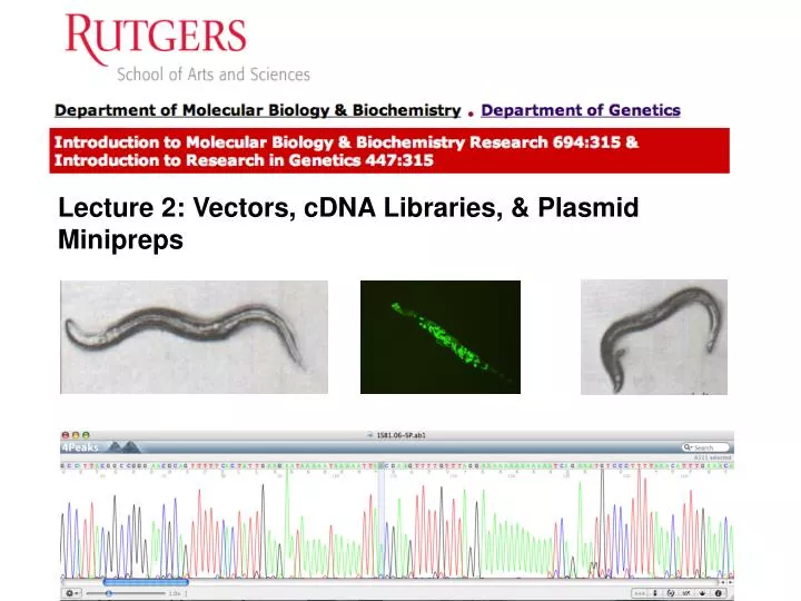
Lecture 2: Vectors, cDNA Libraries, & Plasmid Minipreps. Introduction to Vectors. In order to study a DNA fragment (e.g., a gene), it needs to be amplified and eventually purified. These tasks are accomplished by cloning the DNA into a vector .

E N D
Introduction to Vectors In order to study a DNA fragment (e.g., a gene), it needs to be amplified and eventually purified. These tasks are accomplished by cloning the DNA into a vector. A vector is generally a small, circular DNA molecule that replicates inside a bacterium such as Escherichia coli (can be a virus). p. 2-1
Cloning Scheme Digest Ligate Amplify and Prep p. 2-1
Vector Types There are three commonly used types of vectors: 1) plasmid vectors (e.g., pUC plasmids); 2) bacteriophage vectors (e.g., phage ); and 3) phagemid vectors (e.g., pBlueScriptTM). Each has a different use, and there are many derivatives of these basic building blocks. In this course, you will be using plasmid vectors. p. 2-1
Plasmids • Circular DNA molecules found in bacteria • Replicated by the host’s machinery independently of the genome. This is accomplished by a sequence on the plasmid called ori, for origin of replication. • Some plasmids are present in E. coli at 200-500 copies/cell p. 2-1
Plasmid Engineering • Plasmids also contain selectable markers. • Genes encoding proteins which provide a selection for rapidly and easily finding bacteria containing the plasmid. • Provide resistance to an antibiotic (ampicillin, kanamycin, tetracycline, chloramphenicol, etc.). • Thus, bacteria will grow on medium containing these antibiotics only if the bacteria contain a plasmid with the appropriate selectable marker. p. 2-2
Transforming plasmids into bacteria p. 2-2
Safety Features • Modern cloning plasmids have been engineered so that they are incapable of transfer between bacterial cells • Provide a level of biological containment. • Naturally occurring plasmids with their associated drug resistance genes are responsible for the recent rise in antibiotic-resistant bacteria plaguing modern medicine. p. 2-3
Screening for Inserts p. 2-3
Cloning a DNA fragment into pDONR222 Transform Transform Viable Lethal p. 2-4
DNA Libraries • DNA library - a random collection of DNA fragments from an organism cloned into a vector • Ideally contains at least one copy of every DNA sequence. • Easily maintained in the laboratory • Can be manipulated in various ways to facilitate the isolation of a DNA fragment of interest to a scientist. • Numerous types of libraries exist for various organisms - Genomic and cDNA. p. 2-5
Construction and analysis of a genomic DNA library p. 2-5
Construction of a cDNA library p. 2-6
Differences between a genomic and cDNA library Genomic Library Promoters Introns Intergenic Non-expressed genes cDNA Library Expressed genes Transcription start sites Open reading frames (ORFs) Splice points p. 2-7
Purification of mRNA Collect and grind up animals in mild denaturing solution Spin out debris (Tissue, membranes, etc) Treat with DNAse (removes DNA) Treat with Phenol (removes protein) p. 2-8
Synthesis of cDNA from mRNA p. 2-8
Cloning a DNA fragment into pDONR222 Transform Transform Viable Lethal p. 2-4
Preparing Plasmid DNA • In order to use a vector for cloning, sequencing, etc., it is necessary to isolate the vector in a highly purified form. • Routinely done by most labs. • Many companies now sell “kits” which provide all the solutions necessary for preparing DNA. • Based on similar procedures p. 2-10
Essential components of minipreps • Gentle lysis step to break open the cells and release the plasmid DNA into solution. • Cell debris and chromosomal DNA of the bacteria is pelleted during the centrifugation. • Plasmid DNA remains behind in the clear nonpelleted fraction (the nonpelleted solution left after centrifugation is known as the supernatant). • Subsequent steps are then performed on the supernatant to remove contaminating RNA and proteins from the plasmid DNA. p. 2-10
Grow an overnight (ON) culture of the desired bacteria in 2 ml of LB medium containing the appropriate antibiotic for plasmid selection. Incubate the cultures at 37°C with vigorous shaking. 1. Grow the bacteria p. 2-11
Naming your clones Day Your initials Year T20AV12.09 Group # Clone # T (Teusday), W (Wednesday), H (Thursday)
2a. Transfer the cells to a tube and centrifuge Transfer 1.5 ml of the culture to a microfuge tube and pellet the cells for 1 minute at full speed (12,000 rpm) in the microcentrifuge. First tap or gently vortex the glass culture tube to resuspend the cells which have settled. The culture can be transferred to the microfuge tube by pouring. p. 2-11
2b. Remove the supernatant Remove the growth medium (supernatant or sup) by aspiration or by using the P-1000. Leave the bacterial pellet as dry as possible so that additional solutions are not diluted. p. 2-11 (fig not shown)
3. Resuspend the cell pellet Resuspend the bacterial pellet in 250 µl of Buffer PI by vigorous vortexing. Add 150 ml of PI, cap the tube, and vortex on the highest setting (pipetman can be used). Look very closely for any undispersed pellet before proceeding to the next step. It is essential that the pellet be completely dispersed. PI contains two essential components: Tris and EDTA. Tris is used to buffer the pH of the cell suspension. EDTA is a chemical that chelates divalent cations (ions with charges of +2) in the suspension, such as Mg++. This helps break down the cell membrane and inactivate intracellular enzymes. p. 2-11
4. Add Solution P2 Add 250 µl of Solution P2 (0.2 N NaOH, 1%SDS), mix gently 4-6 times. Do not vortex!!This will shear the DNA and contaminate your DNA preps. Denatures protein, DNA, RNA, membranes. During this step a viscous bacterial lysate forms (the cells lyse). p. 2-12
5. Add Solution N3 • Add 350 µl of Solution N3 (3 M KOAc, pH 4.8). Mix gently 4-6 times.Do not vortex. • P3 neutralizes cell suspension. A white precipitate consisting of aggregated chromosomal DNA, RNA and cell debris and SDS will form. • Plasmids will renature p. 2-12
6. Centrifuge cell debris Centrifuge for 10 minutes at full speed in the microcentrifuge. A white pellet will form on the bottom and side of the tube after centrifugation. p. 2-13
7. Transfer sup. (DNA) to spin column. Using a P-1000 set at 600ul, transfer the supernatant to the appropriately labeled spin column which has been inserted into the 2 ml microcentrifuge tube. p. 2-13 (Fig not shown)
8. Centrifuge the spin column Centrifuge for 1 minute at full speed, and drain the flow-through from the collection tube. p. 2-13
9. Wash the column with PE • Add 750 ul of Wash buffer PE to the spin column contained in the 2 ml Collection Tube, centrifuge at full speed for 1 minute, and drain the flowthrough. • This buffer helps to further remove any nucleases that may have co-purified with the DNA. Remove the liquid that has passed through the column in the same way as performed in Step 9. p. 2-13
10. Spin the column to remove PE • Place the spin column in a fresh 1.7 ml microcentrifuge tube (with lid cut off) and centrifuge again for 1 minute at full speed to remove any residual wash solution that might still be in the column. • Any residual wash solution must be removed because the ethanol contained in this solution may interfere with further DNA manipulations. It is normal to remove a small amount of liquid from the column at this step, however if a significant amount of solution (50-100 ul or greater) is found in the collection tube, repeat this step. p. 2-14 (Figure not shown)
11. Elute the DNA with EB Place the spin column into an appropriately labeled 1.7 ml microcentrifuge tube and add 50 ul of EB buffer to the column.Centrifuge at full speed for 1 minute. Elutes the plasmid DNA from the column and collects in the microcentrifuge tube. p. 2-14
12. Store your DNA Remove the spin column from the labeled 1.7 ml microcentrifuge tube and close the lid on the tube tightly. Store the miniprep DNA in your freezer box (-20C). p. 2-14
Other types of vectors 1. Phage Vectors p. 2-15
2. Phagemids Have plasmid and phage components F1 Phage origin of replication for making single strand DNA p. 2-15
Vector Insert Size Vector Type Cloned DNA (Kb) Plasmid 20 Phage 25 Cosmid 45 BAC 300 YAC 1000 p. 2-17