Download
1 / 20
200 likes | 498 Views
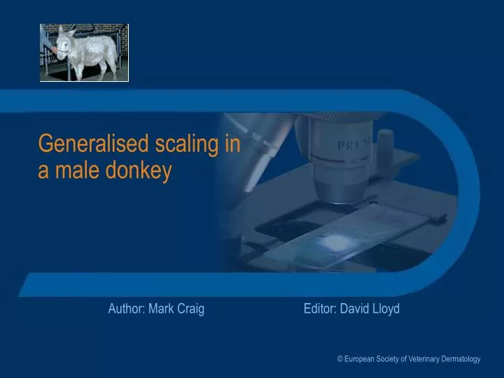
Generalised scaling in a male donkey. Author: Mark Craig. Editor: David Lloyd. © European Society of Veterinary Dermatology. History. 10-year-old entire male donkey First signs Reduced appetite, weight loss, generalised scaling In progress over a 3-month period Treatment by referring vet

E N D
Generalised scaling ina male donkey Author: Mark Craig Editor: David Lloyd © European Society of Veterinary Dermatology
History • 10-year-old entire male donkey • First signs • Reduced appetite, weight loss, generalised scaling • In progress over a 3-month period • Treatment by referring vet • Intramuscular penicillin/streptomycin daily for 10 days • No improvement Click to reveal the text on this screen Click the forward arrow to jump to the next screen History
Clinical signs - 1 • Generalised exfoliative erythroderma • Thin, depressed • Rectal temperature, pulse rate, respiratory rate normal • No peripheral lymphadenopathy • No oral lesions were present Signs
Clinical signs - 2 • The donkey is thin and depressed • There is poor coat with generalised scaling Signs
Clinical signs - 3 Exfoliation and erythema of the scrotum Periocular scaling and greasy matted hair around the eye Signs
Clinical signs - 4 Close up of scaling, matting of coat and underlying erythema Signs
How would youapproach this case? • What are the next steps you would take? • Make a list of your principle differential diagnoses • List any samples you would collect • List any tests you would perform to assist in making a definitive diagnosis Signs
Differential diagnoses • Bacterial infection including dermatophilosis • Dermatophytosis • Pemphigus foliaceus, SLE • Drug eruption • Cutaneous lymphoma Differentials
Tests - 1 • Skin scrapings • Blood tests: routine haematological and biochemical screens • Fungal culture of scale and hairs • Multiple skin biopsy samples Tests
Tests - 2 • Scrapings did not reveal ectoparasites or fungal structures • Scales/crusts were emulsified and smears examined for bacteria including Dermatophilus; no significant findings • Haematology: marked leukocytosis (35.1 x 10³/mm3) with neutrophilia and lymphocytosis, slightly reduced RBC count. • Blood biochemistry: raised total protein, hyperglobulinaemia, raised ALP and CK Tests
What now? • What treatment should you now institute, if any, whilst waiting for the fungal cultures and biopsy results? • What are now your principle differential diagnoses? • Are there any other samples you would collect? Tests
Tests - 3 • No immediate action taken • No parasites or evidence of dermatophytes demonstrated in scrapings • Smears failed to reveal significant bacteria • The leucocytosis (neutrophilia + lymphocytosis) were suggestive of possible bacterial infection but the blood biochemistry results were not diagnostic • Antibacterial therapy might have been instituted but was inhibited by cost and because no significant deterioration was expected before biopsy results were available Tests
Histopathology Results An interface dermatitis pattern predominated, possible indicating lupus or a drug eruption Tests
What is yourdiagnosis? • Do the investigations permit a definitive diagnosis? • Are there any additional investigations which you think may need to be done Tests
Further steps • Consultation with the pathologist • The histological picture was not clear and the pathologist suggested a second opinion supported by immunohistochemical studies Tests
Histopathology Results Another view of the histopathology showing lichenoid infiltration and microabscess formation with predominantly mononuclear cells Tests
Diagnosis • Subsequent immunohistochemical studies showed a strong reaction to CD-3 of infiltrating cells. • A diagnosis of epidermotropic lymphoma was made Tests
How would you deal with this case? • What is your prognosis? • How will you advise the owner? • What treatment would you consider? Therapy
Prognosis • Prognosis is grave • Disease is fatal • Steroids and cytotoxic drugs are unlikely to be helpful • Euthanasia was carried out Therapy
Review • If you would like to review this case, please use the navigation buttons below Notes