Download

1 / 19
250 likes | 1.74k Views
Approaches to the Pineal Region. Jan M. Eckermann, MD Department of Neurosurgery. Why go there?. Pineal cell tumors: pineocytomas, pineoblastomas Germ cell tumors: teratomas, dermoid, epidermoid, endodermal sinus, embryonal cell, choriocarcinoma, germinoma,

E N D
Approaches to the Pineal Region Jan M. Eckermann, MD Department of Neurosurgery
Why go there? • Pineal cell tumors: pineocytomas, pineoblastomas • Germ cell tumors: teratomas, dermoid, epidermoid, endodermal sinus, embryonal cell, choriocarcinoma, germinoma, • Astrocytomas, meningioma, ependymoma, metastatic tumors
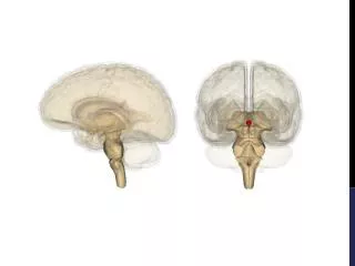
The Pineal Region • Anterior: Quadrigeminal plate, pineal body, habenular complex • Lateral: Mesial temporal and occipital lobes, pulvinar • Roof: Splenium • Floor: Vermis
The Quadrigeminal Cistern • Both supra- and infratentorial • Anterior: Superior medullary velum, quadrigeminal plate, pineal gland • Posterior: Thick arachnoid to tentorium • Lateral: Loose arachnoid separates from ambient cisterns
The Quadrigeminal Cistern • Structures within: • Great vein of Galen • Terminal internal cerebral veins • Basal vein of Rosenthal • Pericallosal veins • Internal occipital veins • PCA (P4) • Posterior choroidal a. cisterna velum interpositum
Approaches • Supracerebellar – Infratentorial • Occiptial – Transtentorial • Combined Supratentorial – Infratentorial Transsinus
Supracerebellar – Infratentorial • Sitting or concord position • Midline or inverted U-shaped incision
Occipital - Transtentorial • Three – quarters prone position • Operative side in dependent position • Inverted J
Combined Supratentorial – Infratentorial Transsinus • Semiprone position • Operative side in dependent position • Inverted J • Craniotomy made in three pieces
Complications and Considerations • Supracerebellar – Infratentorial: • Air embolism • Ventricluar collapse SDH, pneumocephalus • Not suitable for superior extending lesions • Gravity retracting cerebellum
Complications and Considerations • Occiptial – Transtentorial: • Retraction of occipital lobes visual field defects • Disconnection syndrome • Limited exposure of contralateral side • Good view of quadrigeminal plate
Complications and Considerations • Combined Supratentorial – Infratentorial Transsinus: • Brain edema • Venous infarcts • Very wide exposure • Consider primary re-anastomosis or patch graft
References • Fossett TF and Caputy JC. Operative Neurosurgical Anatomy. Thieme: New York 2002 • Haye AH and Laws ER. Brain Tumors. Churchill Livingstone: Edinburgh 1995