Download
1 / 65

E N D
Fascial Layers of the Neck MutasamArifSaeed 3rdstagemedicalstudent
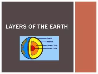
Overview Fascia is an internal connective tissue which forms bands or sheets that surround and support muscles, vessels and nerves in the body. In the neck, these layers of fascia not only act to support internal structures, but also help to compartmentalise structures of the neck. There are two fascias in the neck – the superficial cervical fascia and the deep cervical fascia.
Superficial Cervical Fascia The superficial cervical fascia lies between the dermis and the deep cervical fascia. It contains numerous structures:Neurovascular supply to the skin,Superficial veins (e.g. the external jugular vein),Superficial lymph nodes,FatPlatysma muscle. Subcutaneous cervical tissue--->
Superficial Cervical Fascia Platysma The superficial cervical fascia blends with the ‘paper thin’ platysma muscle. The platysma is a broad superficial muscle which lies anteriorly in the neck. It has two heads, which originate from the fascia of the pectoralis major and deltoid. The fibres from the two heads cross the clavicle, and meet in the midline, fusing with the muscles of the face. Superiorly, the platysma inserts into the inferior border of the mandible. Innervation to the platysma is via the cervical branch of the facial nerve.
Deep Cervical Fascia The deep cervical fascia lies, as its name suggests, ‘deep’ to the superficial fascia and platysma muscle. This fascia is organised into several layers. These layers act like a shirt collar, supporting the structures and vessels of the neck.
layers of the deep cervical fascia (superficial to deep): InvestingLayer The investing layer is the most superficial of the deep cervical fascia. It surrounds all the structures in the neck. Where it meets the trapezius and sternocleidomastoid muscles, it splits into two, completely surrounding them. attachmentsofinvesting fascia: Superior – attaches to the external occipital protuberance and the superior nuchal line of the skull. Anteriorly – attaches to the hyoid bone. Inferiorly – attaches to the spine and acromion of the scapula, the clavicle, and the manubrium of the sternum. Posterior – attaches along the nuchal ligament of the vertebral column
Pretracheal Layer(Visceral fascia) The pretracheal layer of fascia is situated in the anterior neck. It spans between the hyoid bone superiorly and the thorax inferiorly (where it fuses with the pericardium). The trachea, oesophagus, thyroid gland and infrahyoid muscles are enclosed by the pretracheal fascia. Anatomically, it can be divided into two parts: Muscular part – encloses the infrahyoid muscles. Visceral part – encloses the thyroid gland, trachea and oesophagus. The posterior aspect of the visceral fascia is formed by contributions from the buccopharyngeal fascia (a fascial covering of the pharynx).
Prevertebral Layer The prevertebral fascia surrounds the vertebral column and its associated muscles; scalene muscles, prevertebral muscles, and the deep muscles of the back. It has attachments along the antero-posterior and supero-inferior axes: Superior attachment – base of the skull. Anterior attachment – transverse processes and vertebral bodies of the vertebral column. Posterior attachment – along the nuchal ligament of the vertebral column Inferior attachment – fusion with the endothoracic fascia of the ribcage. The anterolateral portion of prevertebral fascia forms the floor of the posterior triangle of the neck. It also surrounds the brachial plexus as it leaves the neck and subclavian artery as it passes through the lower neck region – in doing so, it forms the axillary sheath.
CarotidSheath The carotid sheaths are paired structures on either side of the neck, which enclose an important neurovascular bundle of the neck. The contents of the carotid sheath are:Common carotid artery,Internal jugular vein,Vagus nerve,Accompanying cervical lymph nodes. The fascia of the carotid sheath is formed by contributions from the pretracheal, prevertebral, and investing fascia layers. The carotid artery bifurcates within the sheath into the external and internal carotid arteries. The carotid fascia is organised into a column, which runs between the base of the skull to the thoracic mediastinum. This is of clinical importance as a pathway for the spread of infection.
Clinical Relevance: Fascial Spaces of the Neck The neck fascia compartmentalises structures within the neck. These layers of tough fascia can limit the spread of infection (for example, a superficial skin abscess may be prevented from spreading deeper into the neck by the investing fascia). However, infections that reach the potential spaces between the neck fascia have a well-defined spread: Retropharyngeal space – located between the buccopharyngeal fascia (posterior aspect of the visceral pretracheal fascia) and the prevertebral fascia. Extends from the base of the skull to the posterior mediastinum. Visceral space – enclosed by the visceral pretracheal fascia. Extends from the hyoid bone to the superior mediastinum.