Download

1 / 16
0 likes | 1 Views
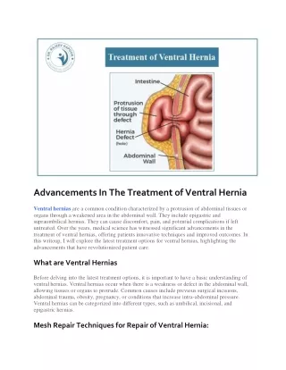
Ventral hernias refer to fascial defects of the anterolateral<br>abdominal wall through which intermittent or continuous<br>protrusion of abdominal tissue or organs may occur<br>(Fig. 1). They are either congenital or acquired. In adults,<br>more than 80% of ventral hernias result from previous<br>surgery, hence the term incisional hernias. They have been<br>reported to occur after 0u201326% of abdominal procedures.<br>Although these hernias mostly become clinically manifest<br>between 2 and 5 years after surgery, studies have shown<br>that the process starts within the first postoperative month.

E N D
Laparoscopic Repair of Ventral H ernia Prof. Dr. R. K. Mishra INTRODUCTION Ventral hernias refer to fascial defects of the anterolateral abdominal wall through which intermittent or continuous protrusion of abdominal tissue or organs may occur (Fig. 1). They are either congenital or acquired. In adults, more than 80% of ventral hernias result from previous surgery, hence the term incisional hernias. They have been reported to occur after 0–26% of abdominal procedures. Although these hernias mostly become clinically manifest between 2 and 5 years after surgery, studies have shown that the process starts within the first postoperative month. They are said to occur as a result of a biomechanical failure of the acute fascial wound coupled with clinically relevant impediments to acute tissue repair and normal support function of the abdominal wall. Historically, incisional hernias have been repaired with either primary suture techniques or placement of a variety of prosthetic materials. Before the 1960s, most ventral hernias were repaired primarily with suture and a few with metallic meshes. Even with some modifications, recurrence rates with the primary suture repair ranged from 24 to 54%. The introduction of polypropylene mesh (PPM) repair by Usher in 1958 opened a new era of tension-free herniorrhaphy. Recurrence rates with prosthetic mesh significantly decreased down to 10–20%. Subsequently, it was realized that the placement and fixation of the mesh was more crucial in determining the outcome of the repair. The concept of placement of the mesh in the preperitoneal, retromuscular position with a wide overlap of at least 5 cm over the hernia defect in all directions was introduced in the late 1980s. The refinement of this method decreased the recurrence rates to as low as 3.5% making it to be declared as the standard of care of ventral hernias. However, implantation of the mesh by open techniques requires wide dissection of soft tissue contributing to an increase in wound infection and wound- related complications. The treatment of ventral hernias (primary and incisional) represents an underappreciated challenge for surgeons. Ventral hernia results from a weakness in the musculoaponeurotic layer of the anterior abdominal wall. This type of hernia has root of development during the embryonic growth such as omphalocele, gastroschisis, and congenital umbilical hernia. Recently, the ventral hernias are reported more due to iatrogenic factors. Even after laparoscopic surgery, if the 10-mm port is not repaired properly, there is always a chance of ventral hernia (incisional hernia) development. Obviously, the initial closure is the most important factor, since faulty technique will universally lead to development of herniation. There are other associated co-morbid conditions, which may encourage the creation of incisional herniation. These include intra-abdominal sepsis or wound infection, morbid obesity, steroid use, previous use of the incision, hematoma formation, and respiratory compromise with increased cough. Other factors include duration of the operation, crossing incisions, ineffective wound drainage, and excessive wound tension. Two other important variables include nutritional aspects as well as presence of cancer, which reduce the overall ability for wound healing and collagen deposition in the wound. The repair of incisional and ventral hernias continues to be a surgical challenge. Reports published in the medical literature indicate 3–13% of laparotomy patients develop incisional hernias. Moreover, clinical studies indicate that the traditional, or open, technique to repair large abdominal wall defects is associated with recurrence rates ranging from 25 to 49%. Fig. 1: Laparoscopic view of ventral hernia.
240 SECTION2: Laparoscopic General Surgical Procedures Among the noniatrogenic ventral hernias, divarication of rectus abdominis, umbilical, paraumbilical, spigelian, and epigastric are more common. In 1992, a successful series of laparoscopic incisional hernia repairs was reported in the medical literature. Since then, the technique has been refined and has grown in acceptance within the surgical community. The laparoscopic technique for ventral hernia repair involves the placement of a tension-free prosthetic bridge across the musculofacial defect rather than attempting to approximate the edge of defect. The hernia defect is covered by appropriate size of mesh, once the content of the sac is reduced. Most of the time, sac content is omentum. Sometime omentum is adhered so strongly that electrosurgical dissection with the help of bipolar is essential. Recently, many newer types of meshes are available in which polytetrafluoroethylene (PTFE) and polypropylene are more popular. There was always a fear of bowel adhesion and fistulization with use of PPM but the clinical evidence of thousands of surgeries has suggested that the omental adhesion is expected but bowel adhesion is not common and intraperitoneal placement of PPM is quite safe. Almost all types of ventral hernia can be repaired by minimal access surgical approach. Hernias like multiple defects (Swiss cheese hernias) are greatly benefited by this approach as all defects get directly visualized and appropriately covered by single mesh. Contraindication of laparoscopic repair of ventral hernia is very large hernia with huge protrusion of skin, which is thin enough. Skin folds mandate correction by abdominoplasty. Dense intra-abdominal adhesions are also a relative contraindication of laparoscopic repair of ventral hernia. Ventral hernias with an associated acute moderate-to-high grade gastrointestinal tract obstruction can be difficult to repair with minimally invasive techniques. Distended bowel can easily be injured with trocar placement or manipulation with laparoscopic instruments, which could further complicate the repair, if mesh is used. Definitive treatment of ventral hernias in patients with an infected mesh and associated complications, such as enteric fistulas, often requires abdominal wall reconstruction with component separation techniques and sublay mesh placement. Intraperitoneal permanent mesh placement is generally not recommended in infected or contaminated fields, and intraperitoneal placement of biologic or bioabsorbable meshes may be associated with significantly increased rates of complications and recurrences. abdominal wall by giving strength. The parietal peritoneum in ventral hernia extends into the defect to form the sac. Adhesions to adjacent viscera must be divided to define the defect. Large ventral hernias with a defect width >8–10 cm or with greater than one-half of the abdominal viscera being outside of the boundary of the abdomen, which is called loss of domain, are difficult to repair without using advanced techniques such as component separation. OPERATIVE PROCEDURES Commonly used techniques: ■ Transabdominal preperitoneal repair (TAPP) ■ Transabdominal retromuscular repair ■ Transabdominal partially extraperitoneal repair (TAPE) ■ Enhanced view totally extraperitoneal repair (eTEP) ■ Endoscopic mini/less open sublay technique/repair (EMILOS) ■ Robotic transabdominal preperitoneal repair (rTAPP) The optimal operative management of primary ventral hernia and incisional hernia is still debatable. No single treatment has been able to tackle all ventral and incisional hernias. LeBlanc and Booth in 1993 first reported application of intraperitoneal onlay mesh (IPOM) for ventral and incisional hernia. It is a relatively straightforward procedure, which in comparison to open mesh repair has been found to reduce chances of surgical site and mesh infection. However, the technique requires expensive fixation devices, which may cause acute and chronic pain. ■ Transabdominal preperitoneal repair: It refers to the laparoscopic ventral hernia and incisional hernia repair whereby mesh is placed in the preperitoneal space, similar to TAPP and total extraperitoneal repair (TEP) for inguinal hernia repair. It involves use of a transabdominal preperitoneal approach and can be used for midline and lateral hernias. ■ The eTEP approach: It has been described previously for inguinal hernia repair. In ventral hernia repair, it relies on initiation of dissection in one retrorectus space and then crossover to the contralateral retrorectus space. The initial port setup and point of crossover depends on the location of defect. Patients with long midline laparotomy scar are a relative contraindication to this technique. ■ Endoscopic mini/less open sublay repair technique: This technique is basically a reversed TEP procedure, which has been designed for midline, epigastric, umbilical, or incisional hernia with coexisting rectal diastasis. It utilizes the original MILOS concept (mini/less open sublay), which has been introduced by Reinpold. A large mesh (20 × 30) is implanted in the retromuscular space via a small skin incision (2–8 cm) without any fixation. ■ Robotic transabdominal preperitoneal repair robotic approach: It is an emerging minimal access technique, LAPAROSCOPIC ANATOMY Ventral hernia develops due to structural weakness of the abdominal wall. The muscle and fascia that span the space between costal margin superiorly, the spine and muscles of the back posteriorly, and the pelvis inferiorly support the
241 CHAPTER17: Laparoscopic Repair of Ventral Hernia which utilizes set principles of open as well as conventional laparoscopic techniques. The popularity of this technique is growing in the west, attributed to its enhanced precision, 3D vision, and surgeon ergonomics. The robotic platform allows exploration of the individual abdominal wall layers and subsequent mesh placement in a preperitoneal, retromuscular, and onlay position. above umbilicus, camera operator should stand left to the surgeon. Monitor should be placed opposite to surgeon and instrument trolley should be toward the leg of the patient. Port Position Technique of laparoscopic repair of ventral hernia is quite simple. First pneumoperitoneum is created at a site away from the defect. Three-port techniques are used for laparoscopic repair of ventral hernia. The first step in performing laparoscopic repair of ventral hernia is gaining access to the free peritoneal cavity. A site distant from any prior incision and the hernia defect is chosen. Typically, this is in the right upper quadrant (RUQ) or left upper quadrant (LUQ) (Figs. 3 to 7). The absence of incisions in these locations does not necessarily guarantee the absence of adhesions to viscera. While many approaches for access to the peritoneal cavity have been described, including blind insufflation and specialty trocars, open access in the fashion of Hasson is by far the safest alternative. Once the pneumoperitoneum is created, all other ports are placed according to baseball diamond concept. The most preferred site of access is left hypochondrium in most midline and lower abdominal defects. Most of the surgeons prefer optical trocar in the left subcostal location, lateral to the midclavicular line (Palmer’s point). We generally go over the costal margin called Mishra’s point and during access, pull the abdominal wall caudally to bring it subcostal (Fig. 4). First access should be preferably through left hypochondria, if Veress needle technique is used and then two other ports should be made so that proper triangle is formed. The distance between two ports should not be less than 5 cm (Fig. 3). The telescope will first enter through left hypo chondriac port but once dissection starts, the telescope will come in the middle, so that the angle between two working ports will become 60°. The 10-mm 30° telescope is better to view anterior abdominal wall (Fig. 7). Achieving free access to the peritoneal cavity represents the greatest risk to the patient. For most ventral incisional Patient Position Acutely incarcerated or strangulated ventral hernias require urgent repair. Chronic symptomatic ventral hernias may be repaired electively assuming there are no medical contraindications. Chronic ventral hernias with minimal or no symptoms may be watched, if the patient wishes to avoid surgical repair. Patient should be clearly informed that laparoscopic repairs will not help cosmetically, if the skin is lax and hanging loosely. Bowel preparation is a good practice to have more room inside the abdominal cavity to handle instruments. After anesthesia, nasogastric tube is required to deflate the stomach completely because in most of the cases, access made through left hypochondrium. Splenohepatomegaly is absolute contraindication of the access through left hypochondrium. In general, both arms should be tucked with all pressure points padded. Patients should be secured to the bed with straps to allow the bed to be steeply tilted into reverse Trendelenburg or Trendelenburg positions when necessary, depending on the location of the hernia. Position of Surgical Team Surgeon stands left to the patient with camera operator on his left or right side depending upon the location of ventral hernia (Fig. 2). If ventral hernia is below the umbilicus, the camera operator stands right to the surgeon. If the defect is Fig. 2: Operating room setup in ventral hernia repair. Fig. 3: Palmer’s point is used for access.
242 SECTION2: Laparoscopic General Surgical Procedures Fig. 4: Veress needle introduction through Palmer’s point. Fig. 5: Palmer’s point is used for trocar introduction. Fig. 6: Telescope is introduced to check any adhesion. Fig. 7: Secondary ports are introduced. hernia repairs, adhesiolysis is required to make room for subsequent mesh insertion, manipulation, and fixation. The difficulty of adhesiolysis is unpredictable, and the presence of PPM should be a red flag indicating the potential for the presence of dense and difficult to dissect adhesions, often involving the bowel. All maneuvers performed as part of the lysis of adhesions must be done under direct vision. This is best carried out by sharp dissection utilizing bimanual palpation of the anterior abdominal wall, placing the adhesions under variable degrees of tension (Figs. 8 and 9). There is significant risk in extensive blunt dissection, as the bowel may be fixed at several points placing it at risk for unrecognized perforation with the tip of dissecting instruments. In spite of the enthusiasm for different energy sources, these are best avoided. As in open cases, dissection should be carried out at the avascular junction of the adhesions and the anterior abdominal wall. Ligating clips or the limited application of an energy source can be used when significant bleeding from vessels is encountered. In the majority of cases, even this is unnecessary. The risk of monopolar cautery is well known, but there is also risk of thermal injury by direct contact with ultrasonic or radio frequency dissection instruments. Ultrasonic dissector should be preferred over monopolar. If bowel is incarcerated, cold scissor dissection should be preferred rather than energy. This is particularly true in the poorly visualized area behind adhesions. If omentum is adhered with the anterior abdominal wall, it should be dissected after applying bipolar or extracorporeal knot (Figs. 10A and B). It is critical that all adhesions to the anterior abdominal wall be released to allow adequate patch placement and fixation. Once adhesiolysis has been completed, the exact extent of the defect can be directly evaluated. The defects are carefully drawn onto the skin of the anterior abdominal (Figs. 11A to C). In the case of multiple defects, the area drawn should include all of the defects. We have transformed now to repairing the entire area of a previous incision as opposed to simply repairing a single defect. There have been a number of patients who have presented later in follow-up and are discovered to have a new hernia, outside the area of previous repair. In open surgery, these may have unknowingly simply been considered recurrences. If there is any difficulty in delineating the margins of the defect, a spinal needle can be passed perpendicular to the anterior abdominal wall and through the margins of the defect. The selection of size of mesh is important to prevent recurrence of hernia and it should be sufficiently big so that approximately 4 cm healthy margin of defect of hernia should be covered all around. Recently, new hybrid mesh has been introduced with absorbable material on one side and unabsorbable prolene on the other. These meshes are better than prolene because adhesions are less likely to develop due to absorbable material toward the bowel.
243 CHAPTER17: Laparoscopic Repair of Ventral Hernia B A D C Figs. 8A to D: Any adhesion should be removed with bipolar and scissors. in a trial of umbilical hernia repairs, this technique should be avoided until further studies demonstrate safety and efficacy. After fixing the mesh, greater omentum is spread like an apron in-between the bowel and mesh (Figs. 12A and B). Some adhesions of mesh with omentum are always expected in this technique. Very high epigastric hernias close to the costal margin are best managed by leaving an overhang of mesh draped over the diaphragm without fixation. Fixation with tacks to the diaphragm should be avoided, as it has been associated with serious complications such as lung and cardiac injuries (tamponade). If patient is mobilized early and newer generations of meshes are used, the long-term complication of adhesions is very less with this technique. Loosely held mesh hanging through the anterior abdominal wall will definitely increase the chances of adhesions with bowel. Tacker should be used to fix the mesh in position. Recently, a technique of using prolene suture to fix the mesh with anterior abdominal wall is being used with the help of suture passer or looping technique with the help of Veress needle cannula (transfascial suture). The main idea of this method is to reduce the cost of surgery, but there is increased chance of infection and adhesions with this method. We also lack any long-term randomized controlled trial (RCT) to prove the outcome of this external suture technique to fix the mesh in ventral hernia repair. Meshes can be fixated to the anterior abdominal wall with transfascial sutures, tacks, or a combination of the two. If no transfascial sutures are placed, two rows of permanent tacks Fig. 9: Bipolar can be used safely in case of adhesion with omentum. The mesh is fixed along margins and around the ring of defect of rectus to ensure a close approximation of mesh to abdominal wall. Care should be taken that mesh should not be corrugated and it should be in proper contact with anterior abdominal wall. Tacker, Protack, or Endo anchor can be used to fix the mesh. Fibrin glue has been used to fix the mesh, but less frequently. Because of a higher-than-expected recurrence rate at 1 year (26% fibrin sealant versus 6% tacks)
244 SECTION2: Laparoscopic General Surgical Procedures A B Figs. 10A and B: Extracorporeal knot can be used in case of adhesion with omentum. A B C Figs. 11A to C: (A)Appropriate size of mesh should be used to prevent recurrence; (B) Transfascial fixation of the mesh with suture inside view; (C) Transfascial fixation of the mesh outside view. A B Figs. 12A and B: Fixation of mesh can be accomplished outer and inner crowning.
245 CHAPTER17: Laparoscopic Repair of Ventral Hernia should be used to fixate the mesh; if transfascial sutures are used, one or two rows of either absorbable or permanent tacks may be placed at the surgeon's discretion. To place transfascial sutures, multiple small “stab” incisions are made in the skin at locations that correspond to those of the previously placed sutures on the mesh (Figs. 11B and C). A transcutaneous laparoscopic suture- passing device is advanced through the stab wound into the peritoneum. The assistant uses a laparoscopic grasper to feed each end of the sutures into the jaws of the awaiting suture passing device, which, when closed, brings the suture through the abdominal wall. Once all the sutures are brought through the abdominal wall, the peritoneal cavity is desufflated and the sutures are tied. The peritoneal cavity is then reinsufflated to a lower pressure (8 mm Hg), and the mesh is examined laparoscopically to ensure appropriate coverage of fascial defect as well as appropriate apposition to the abdominal wall. The mesh is then additionally fixated utilizing single or double rows (outer and inner crowning) of absorbable or permanent tacks, per surgeon preference. If transfascial sutures are not utilized, the stay stitch on the mesh is generally brought through the abdominal wall at the center of the hernia fascial defect to facilitate mesh fixation with a double crown of permanent tacks. Other commercially available mesh positioning devices are available but may be difficult to place. the mesh would not unravel when it was cut. PPM gained widespread popularity over the next 30 years and several types of PPM are commercially available today. Polyester mesh was also introduced in the 1950s in Europe. Rives and Stoppa employed polyester mesh in their landmark article describing a preperitoneal technique of ventral hernia repair in 1989. The technique described by Rives and Stoppa has become the standard by which all abdominal wall incisional hernia repairs are measured. PPM and polyester mesh revolutionized abdominal wall repair because the meshes did not deteriorate with age, were pliable, and would stretch, allowing for more even load distribution. Nevertheless, the large interstices in polypropylene and polyester mesh promoted adhesion formation when the mesh came into contact with the visceral abdominal cavity. Reported complications included small bowel obstruction, erosion, and fistulization. Expanded polytetrafluoroethylene (ePTFE), initially used as a vascular prosthesis, was adapted for abdominal wall incisional hernia repair in 1983 by WL Gore and Associates and modified several times in the 1990s. Unlike the polypropylene and polyester meshes that preceded it, ePTFE is microporous and select products are uniquely designed with pores measuring 3 μ on the visceral side facing the abdominal cavity and 22 μ facing the abdominal wall. This design promotes fibroblastic and vascular ingrowth from the abdominal wall 22 µ side, but inhibits tissue attachment to the material on 3 µ side when exposed to the intra-abdominal cavity. There are no reports of fistulization or small bowel obstructions due to adhesions from ePTFE material. Synthetic meshes, made of materials such as ePTFE and polypropylene, are used most of the time for repair of hernia. The repair process for these materials is based on scar formation in and around the mesh. The advantage of using these materials is that they generally do not react with the human tissue. They are strong and do not tear easily, are readily available, inexpensive, and have a long history of being used for soft tissue replacements. However, use of synthetic materials is not without problems. As a foreign material, body may react to its presence by growing around it (encapsulation) in an attempt to exclude it from body. In the process, tissue forms a capsule of rigid, fibrous scar tissue around the synthetic material. The rigid capsule could affect the function and the esthetic outcome of the repair. Furthermore, foreign bodies such as synthetic materials increase the risk of infection when implanted in the body. As part of the foreign body response, the repair site may be subjected to inflammation, infection, and pain. CHOICE OF MESH IN VENTRAL HERNIA Synthetic Materials A variety of synthetic polymeric meshes were developed in the second half of the 20th century, which revolutionized the hernia repair. Meshes can be categorized in terms of weight, pore size, material, fiber type, and flexibility. Heavyweight meshes tend to form a dense scar plate and are best suited to applications where mechanical stability is a factor. Lightweight meshes are formed from thin fibers and are designed to flex with normal physiological movement. They form a flexible scar and may cause less discomfort than heavyweight meshes. Fibers may be monofilament or multifilament, and the gaps between the fibers, known as pores, can vary depending on the design. In general, a smaller pore size reduces the ability of the mesh to be incorporated into the body’s own tissues, which may a desirable quality, if the mesh is to be used around delicate bowel tissue, to avoid unwanted adhesions. Meshes of the same material may differ between manufacturers in terms of weight, flexibility, shrinkage, and potential for adhesion formation. With these meshes, abdominal wall defects could be repaired without undue tension on the sutured tissue, decreasing the high recurrence rates of abdominal wall hernia repair. Sir Francis Usher introduced woven monofilament PPM in 1958. It was modified to a knitted mesh in 1962 so that Surgisis It is porcine intestinal submucosa and specifically designed as a surgical graft for hernia and abdominal body wall
246 SECTION2: Laparoscopic General Surgical Procedures Fig. 13: Surgisis. Fig. 14: AlloDerm. repair (Fig. 13). Surgisis® GoldTM combines strength with flexibility in a naturally occurring graft material that allows for hernia repair without the need for a permanent synthetic prosthesis. Surgisis® GoldTM supports the surgical site while the body's natural healing process replaces the graft with new host tissue. It is collagen biomatrix, naturally occurring and acellular with 18 months shelf-life. AlloDerm It is biological dermal matrix from processed donated human tissue (Fig. 14). AlloDerm is processed from donated human skin. The tissue goes through a cell removal process while retaining the important biochemical and structural components. AlloDerm is, thus, acellular human tissue. Since AlloDerm is derived from human tissue, there may be a concern that it might harbor disease-carrying viruses. Tissue donors are screened and tested for transmissible diseases including HIV, hepatitis, and syphilis before tissue processing. AlloDerm has been utilized in more than 750,000 implants and grafts to date, without any reported incidence of viral disease trans mission to a patient. AlloDerm repairs damaged tissue by providing a foundation for new tissue regeneration. The skin components preserved in AlloDerm contain the information that will help your own tissue to grow into the AlloDerm after placement at the repair site. Fig. 15: Proceed mesh. Titanized mesh is a lightweight, monofilamentous PPM with a weight of 45 g/m2 and a pore size of 3.0 mm. The polypropylene filaments are coated with titanium dioxide. In the tetanized mesh, the polypropylene filaments are coated with a few layers of atomic titanium using what is known as the PACVD (plasma-activated chemical vapor deposition) technique (Fig. 11). Polyurethane-covered Mesh Polyurethane-coated mesh has a permeable peritoneal side (white color) in polyester for excellent fibroblast coloni- zation and rapid tissue fixation; and a nonabsorbable and nonadherent (blue color) smooth side in polyurethane allowing fluid transfer and contact with viscera. The dual- side mesh is equipped with sutures and has a visual mark printed on polyester for easy transfascial mesh centering in IPOM inguinal hernia surgery. Proceed Mesh It is soft PPM covered with polydioxanone (PDS) and oxidized regenerated cellulose fabric (Fig. 15). Titanized Polypropylene Mesh Despite the vast selection of mesh for hernia repair available, nearly all synthetic meshes for hernia surgery continue to use one or other of three basic materials—polypropylene, polyester, and ePTFE. These are used in combination with each other or with a range of additional materials such as titanium, omega-3, monocryl, PVDF, and hyaluronate. Biological Mesh Biological meshes, derived from animal tissues such as porcine skin or bovine pericardium, can be a useful
247 CHAPTER17: Laparoscopic Repair of Ventral Hernia EXTENDED TOTALLY EXTRAPERITONEAL REPAIR FOR VENTRAL HERNIAS Enhanced view TEP approaches for laparoscopic repairs of incisional and ventral hernias have been described in many recent case series. Extended totally extraperitoneal repair is a novel technique that was first introduced by Jorge Daes in 2012 to address difficult inguinal hernias. The principle is to create a larger space than what is done in TEP to tackle large groin hernias. Purported advantages include the ability to place wide pieces of mesh in the retrorectus/retromuscular spaces. It may also help to facilitate minimally invasive primary fascial closure in the cases of large fascial defects requiring posterior (transversus abdominis) component release techniques. alter native to synthetic fabrics under certain circumstances. Such products are expensive, and can stretch out over time, but offer significantly reduced susceptibility to infection relative to synthetic products. A fully synthetic long-term absorbable mesh made from a combination of fast and slow absorbing fibers has also been developed. The basic concept behind the development of these types of materials is that— they provide proper environment for the population of native cells, generation of connective tissue which ultimately leads to the replacement of defective tissue present in the hernia defect. Although biological meshes show great promise in repairing hernia, currently surgeons are hesitant to opt for this over synthetic mesh materials. Because the connective tissue formed by these materials is only 70–80% strong, emphasizing the inherent defect of their native tissue. Therefore, there is a greater chance for the recurrence of hernia upon usage of these meshes. COMPLICATIONS ■ Bowel adhesion ■ Fistulization ■ Nerve injury ■ Vascular injury ■ Seroma ■ Infection and cellulitis ■ Recurrence ■ Pain Multiple studies have documented that open hernioplasty has significant morbidity. Leber reported a 27% long-term complication rate with open repair, among them being infection, hematoma and seroma, chronic sinus tract formation, mesh extrusion, fistula formation as well as soft tissue problems such as nonhealing wound. White reported 34% of 250 open ventral hernia repairs had wound-related complications. The complications of the open repair mainly relate to the type of mesh that is most commonly used (polypropylene and polyester meshes). In addition, the wide dissection of soft tissue that is required for a Stoppa-type retrorectus repair or a Chevrel-type anterior repair leads to the many wound-related problems. Some patients will develop a fluid collection, what is commonly called a seroma, between the mesh and the abdominal wall. Many of these are not apparent to the patient or the surgeon but some are evident and can be bothersome to the patient. Complications from these seromas have been reported in many studies. Most surgeons do not aspirate these fluid collections for fear of infecting the prosthesis. However, the author has freely aspirated the seromas, if they are large or if they are bothersome to the patient. The author has never seen an infection of the prosthesis from aspiration of these fluid collections if full aseptic precautions are taken. Probably, the most dreaded complication that has been seen is bowel injury. Enterotomy is a well-documented complication and commonly occurs and can be readily visualized and handled through an incision. Laparoscopy presents a whole new situation with respect to enterotomy. PREPERITONEAL REPAIR OF VENTRAL HERNIA This technique is also called inlay technique of laparo- scopic ventral hernia repair. The peritoneum is incised and preperitoneal space is created in this technique. The sac of the hernia is excised nicely. It can give some extra flap of peritoneum for successful overlapping of both the edges. In this technique, margin of defect is sutured intracorporeally to decrease the gap. Pressing the abdominal wall on both the side from outside will help to obliterate the space in this hernia surgery. Either intracorporeal stitches or external mattress sutures can be used to fix the mesh to the musculofacial defect. Once the mesh is fixed, the peritoneum is sutured using Vicryl to cover the mesh. This method of laparoscopic ventral hernia repair (LVHR) is same as that of open surgery and it is supposed that formation of adhesion is less. MINIMALLY INVASIVE COMPONENT SEPARATION In laparoscopic ventral hernia repair, it is often difficult to achieve primary fascial closure in patients with large fascial defects (width >8–12 cm) without a relaxing incision. In open ventral hernia repair, component separation is the most commonly used technique to achieve midline fascial closure. Laparoscopic and robotic-assisted component separation techniques have been described, which can be used to facilitate primary fascial closure in laparoscopic ventral hernia repair. The increasing popularity of minimally invasive techniques, particularly robotic techniques, for abdominal wall reconstruction has led to increased use of robotic posterior component separation techniques. The complication rates of minimally invasive component separation techniques are more challenging than those of traditional open component separation techniques.
248 SECTION2: Laparoscopic General Surgical Procedures Prevention is the first line of defense. Lysis of adhesions is well visualized due to the magnification and high-intensity light source inherent in the laparoscopic technique. It is very important that energy sources be used very sparingly if at all during lysis of adhesions. If a surgeon enters the proper planes, there is very little bleeding and thus low need for energy sources. Inappropriate use of energy sources is a common cause of unrecognized enterotomy. Monopolar cautery has the problem of current spread, and it is easy to coagulate one area and see the current spread to the adjacent area instantaneously. For this reason, monopolar cautery should not be used adjacent to the bowel. The ultrasonic or radio frequency dissection instruments are “sold” with the supposed advantage that there is minimal thermal spread unlike monopolar cautery. Although this may be true, the tip remains very hot and any touching of viscera can cause a burn that may not be apparent during the operation. It is only after several hours, either that night or the next day, when the tissue sloughs, that is when the enterotomy presents itself. We do not recommend the use of ultrasonic or radio frequency dissection instruments for this reason. The most important thing to remember is that if lysis of adhesions involving the intestine is not safe, i.e., the surgeon cannot see well or the surgeon cannot determine if an enterotomy has occurred, the patient should be opened! Deaths have been reported from laparoscopic incisional hernioplasty due to bowel injuries that have not been perceived during surgery and only become apparent postoperatively. By the time, the diagnosis is made, the patients are generally septic, and may succumb to this complication. to have shorter operating time depending on the surgeon’s experience, shorter hospital stay, and lower complication rates especially wound and mesh infections and lower recurrence rate during the follow-up period. This evidence has led to the suggestion that now; it would be unethical to conduct a prospective RCT comparing LVHR and open approach. Laparoscopic ventral hernia repair techniques are based on the fundamental principles of the open preperitoneal repair described by Stoppa and Rives. The placement of a large mesh in the preperitoneal location allows for an even distribution of forces along the surface area of the mesh, which may account for the strength of the repair and the decreased recurrence associated with it. The repair capitalizes on the physics of Pascal’s principle of hydrostatics by using the forces that create the hernia defect to hold the mesh in place. For this, to attain maximum effect, there has to be a wide mesh overlap over the defect and adequate, secure fixation. In the open approach, attaining an overlap of 3–5 cm requires extensive soft tissue dissection, with the resultant increase in wound complications. Larger defects should require more overlap and smaller ones theoretically less. The laparoscopic approach not only allows clear definition of the defect margins but also the identification of additional defects that may not have been clinically apparent preoperatively. Both the inlay and onlay placements of prosthetic mesh embrace these fundamental principles of hernia repair. The onlay and the transabdominal inlay methods allow for adequate diagnostic laparoscopy to clearly define the margins and the number of the hernia defects including the occult ones. DISCUSSION Initially described in 1992, laparoscopic repair of incisional hernias has evolved from an investigational procedure to one that can safely and successfully be used to repair ventral hernias. The well-established benefits of laparoscopy repair are less postoperative pain, reduced hospital stay and recovery time, low complication and recurrence rates based on numerous reports, meta-analysis, and few randomized trials. Conventionally, the LVHR entails the intraperitoneal placement and fixation of the prosthetic mesh. Many alternative techniques have been tried in few studies and propose to be an advancement of the conventional approach. Despite its significant prevalence and associated morbidity, there is little in the way of evidence-based guidelines regarding the timing and method of repair of ventral and particularly, incisional hernias. Several large studies on LVHR have been reported. This technique has proven to be a safe and feasible alternative to open mesh repair. Although many are retrospective series and a few comparative studies, only two completed randomized trials comparing open versus laparoscopic mesh repair have been published. Based on these studies, LVHR has been found MESH PLACEMENT AND FIXATION One of the critical technical points that significantly impacts on any method of hernia mesh repair is adequate mesh fixation. The mesh is held in position by sutures and/or staples, clips, tacks, intra-abdominal pressure, and later by fibrinous growth. The most widespread technique in onlay approach involves fixation of mesh with tacks and transabdominal permanent sutures. Some surgeons have tried to reduce the operating and possibly postoperative discomfort by reducing or eliminating the use of sutures. The physics of mesh fixation does not support the sole placement of tacks. Majority of the meshes used are about 1-mm thick. A perfectly placed tack can be expected to penetrate only 2 mm beyond the mesh thus tacks will not give the same holding strength as full-thickness abdominal wall suture. Furthermore, the mesh is placed against the peritoneum, so any ingrowth is most likely into the peritoneum and not into the fascia. Detachment of tacks has also been an attributing factor to some recurrence of hernias. Postoperative recurrence of
249 CHAPTER17: Laparoscopic Repair of Ventral Hernia ventral hernia repair is reported to be as high as 13% when only a stapling, clipping, or the tacking device is used for mesh fixation. Proper use of the transfascial fixation sutures in combination with staples decreased the recurrence rate to as low as 2%. Therefore, the current recommendation for mesh fixation is that a transfascial suture should be placed at a distance of 5 cm each along the perimeter of the mesh and tacking devices be used to affix the edge of the mesh at 1-cm intervals. The preperitoneal approach mesh fixation differs in that, there is immediate and continued fixation by the intact peritoneal sac and whether tacks or sutures or both are used, they fix the mesh directly onto the fascia. The primary concern of the peritoneal flap in the inlay technique is to achieve secure fixation of the mesh to the underlying fascia. The fibrinous ingrowth is from the fascia and not the peritoneum. Furthermore, the preperitoneal positioning confers with the original design of Stoppa. Perhaps the most compelling advantage of the preperitoneal placement of the mesh in the inlay approach is the avoidance of direct interaction between the mesh and the intra-abdominal viscera. Contact of the viscera with foreign material such as the prosthesis may lead to an inflammatory response and adhesion formation, which can induce chronic pain, intestinal obstruction, enterocutaneous fistula, and infertility. In addition, adhesions complicate any future intra-abdominal surgery. The peritoneal covering also allows the use of conventional meshes, which have been associated with intense inflammatory response and adhesion formation by some workers. The choice of the mesh used in LVHR may be the most contentious issue, particularly when financial cost is a major consideration. The biomaterials available for ventral hernia repair have undergone many changes over the last several years. There are new products that have either been recently introduced or are in developmental stages. All seek to achieve two goals—rapid and permanent ingrowth into the body wall and diminution of the risk of intestinal adhesions while maintaining its tensile strength. The visceral side should be smooth, nonerosive antiadhesive, and not easily susceptible to infection. This visceral barrier should be present for at least 1 week because this is the time frame in which adhesions form. The ventral side should be macroporous allowing for fibroblast in growth and a foreign body reaction may be necessary for incorporation and high-tensile strength. Polypropylene (prolene) mesh, introduced by Sir Francis Usher in 1958 and modified in 1962, has gained widespread popularity and several types are commercially available today. Polyester mesh was introduced in Europe in the 1950s. Stoppa used the polyester mesh in their landmark article describing preperitoneal repair of ventral hernia in 1989. Prolene mesh is currently the most widely used because it is relatively inexpensive, easy to handle, has a memory (allows them to regain the original shape), and is firmly incorporated in the abdominal wall due to its ability to induce an intense inflammatory reaction. A 2–5% fistula rate has been reported with PPM used intra-abdominally leading to the suggestion that great care must be taken to separate it from the bowel, if it has to be used at all. However, some studies do not support this view. Bingener found no association of visceral adhesion when prolene was used with adequate omental interposition between it and the bowel. In another study involving 136 patients, Vrijland concluded that enterocutaneous fistula appears to be very rare after prolene mesh repair regardless of intraperitoneal placement, omental coverage, or closing the peritoneum. A study comparing the biomaterials used in LVHR found polyester to have the highest incidence of infection, fistulization, and recurrence. The ePTFE has the longest history in the use for these hernias repair. The original description of the procedure used an early generation of the ePTFE product. The current product has one smooth surface with 3 µ ePTFE interstices, while the other side has 22 µ interstices to facilitate fibroblastic ingrowth for firm fixation. Other modifications of this product involve incorporation of antimicrobials on the visceral surface. All of the composite prostheses have ePTFE and prolene or polyester but differ in the number and attachment of them together. There are no reports of intestinal fistulization or obstruction with ePTFE though it has also been found to induce inflammation and fibrosis in laboratory animals. However, the use of synthetic materials is not without problems. As a foreign material, the repair site is subjected to inflammation, susceptibility to infection and pain as a foreign body response. Encapsulation could affect the elastic function of the abdominal wall and esthetic outcome of the repair. This has stimulated the search for natural biological prostheses such as surgisis, collagen, glycosaminoglycans from porcine intestinal submucosa, and AlloDerm. The financial cost to clinical benefit ratio for use of the substantially expensive composite meshes is unquantified and is likely to remain, as such because, given the widespread acceptance of composite products, a randomized, clinical comparison with prolene is unlikely to occur. Therefore, in selected circumstances, it may be acceptable to use a simple mesh, if this can be excluded from the bowel by tissue interposition be it omentum or peritoneum. A composite mesh should be considered as the current standard of care. The extraperitoneal placement of the prostheses would in principle diminish the intra-abdominal complications associated with formation of adhesions. It would also allow the safe use of the conventional meshes such as prolene, which has high intrinsic tensile strength, good memory, and is cheaper. In addition, the peritoneal coverage over the entire mesh provides additional security of fixation and a better mechanical advantage. As such, it can be seen as an advance over the onlay approach. However, the placement is
250 SECTION2: Laparoscopic General Surgical Procedures technically demanding as evidenced by the high-iatrogenic peritoneal tears in the largest series and it may not be feasible in the scarred abdomen of incisional and recurrent hernias, which constitute the bulk and seems to benefit most, from LVHR. Thus, the issue of limitation of patient population among the technical feasibility and adequacy of defect coverage are issues of great concern before the method is accepted as an additional procedure for LVHR. The good results and the attributed safety of LVHR are based on the large number of studies mainly utilizing the intraperitoneal approach. The generalization of the procedure has resulted in multiple variations of techniques. Overall, fewer complications are reported after LVHR than after open mesh repair, especially in relation to wound and mesh infection. The efficacy of the inlay approach as an advancement of the conventional repair needs to be evaluated in terms of the several specific complications that are of particular relevance in laparoscopic procedures. Fig. 16: Adhesiolysis for adhesion developed during previous intraperitoneal onlay mesh (IPOM). has been released and freed of its attachments. Once the injury is recognized, it is the surgeon’s level of comfort with laparoscopic suture repair that determines the best approach. With minimal spillage of bowel contents, the injury may be treated with either laparoscopic repair or open repair; the latter usually can be carried out through a mini-laparotomy over the injured area. Whether the mesh prosthesis is put primarily or later depends on the degree of contamination. More significant bowel injuries necessitate conversion to open repair. Missed injuries manifest postoperatively mandating re-exploration with occasional removal of the mesh and immediate recurrence of the hernia. BOWEL INJURY Probably the most dreaded complication is bowel injury, and particularly if it is missed intraoperatively. It is a potentially lethal complication. The overall incidence of bowel injury does not differ significantly between open repair and laparoscopic repair and is generally low with either approach (1–5% when serosal injuries are included). Pneumoperitoneum may hinder the recognition of bowel injury at the time of operation. There have also been reports of late bowel perforation secondary to thermal injury with laparoscopic repair. Minimizing the use of electrocauterization and ultrasonic dissection markedly reduces the risk of bowel injury. The visualization afforded by the pneumoperitoneum places adhesions between the abdominal wall and the bowel under tension. The high-intensity light source and the magnification inherent in the laparoscopy facilitate identification of the least vascularized planes. As far as possible, direct grasping, the bowel should be avoided preferring simply to push it or to grasp the adhesions themselves to provide countertraction. External pressure on the hernia may also help. Larger vessels in the omentum or adhesions are controlled with clips. Some degree of oozing from the dissected areas is tolerated; such oozing almost always settles down without specific hemostatic measures. CELLULITIS Compared with open ventral hernia repair, laparoscopic repair causes fewer surgical site infections, both superficial and deep. About 2–4% of patients develop abdominal wall cellulitis after laparoscopic ventral hernia repair as a result of either infection or inflammatory response to the prosthetic mesh. Postoperative cellulitis can be treated with antibiotics. SEROMA Seroma formation is one of the most commonly reported complications in LVHR, though it is not unique to laparoscopy (Figs. 17A and B). It is a swelling-like mass that occurs immediately after operation in virtually all patients, but disappears after a few weeks. Most seromas develop above the mesh and within the retained hernia sac. The mean incidence of seroma in reported series at a range of 4–8 weeks is 11.4%. In the largest multi-institutional trial, seromas that were clinically apparent for more than 8 weeks were considered a complication and occurred in 2.6% cases. Regardless of whether they are aspirated under sterile conditions or allowed to resolve, they rarely cause long-term morbidity. Aspiration may increase the risk ADHESION (FIG. 16) In cases of dense adhesions, it is preferable to divide the sac or the fascia rather than risk injury to bowel (Fig. 1). Densely, adherent PPM is best excised along with the abdominal wall rather than attempting to separate it from the serosa of the bowel. If bowel injury is suspected, immediate and thorough inspection should be carried out. It may be difficult or impossible to find the exact site of injury later once the bowel
251 CHAPTER17: Laparoscopic Repair of Ventral Hernia A B Figs. 17A and B: Seroma developed after ventral hernia repair. of mesh infection but is recommended if they enlarge or persist before they reach their extremes. Patients sometimes mistake a tense seroma for recurring incisional hernia, but appropriate preoperative discussion should provide them with significant reassurance on this point. Although Feldman suggests that seroma formation is not related to a particular type of mesh, Carbanjo and Heniford reported a higher incidence of seroma formation with ePTFE than prolene-based meshes. The low incidence in the latter meshes has been attributed to the large pores of the prolene-based meshes that allow more efficient resorption of wound secretions into the abdominal cavity than ePTFE meshes. The dissection of the preperitoneal space during the inlay method may lead to more seroma formation. This is supported by the fact that—in the classical description of the onlay technique, it is emphasized that no attempts should be made to reduce or resect the hernia sac. This has been established to be unnecessary and to increase the incidence of seroma formation. The peritoneum interposed barrier between the mesh and the abdominal cavity may hinder the direct drainage of this fluid regardless of the mesh used. Thus based on these facts, it seems plausible that the problem of seroma formation is expected to be higher in the inlay than the conventional onlay approach. is a grave concern in LVHR, particularly after a difficult adhesiolysis, correct interpretation of the significance of postoperative pain is an important issue. Whether or not to relaparoscope a patient who experiences severe pain remains an important issue. A possible explanation of the common type of pain may be that—the transabdominal suture entraps an intercostal nerve, as it courses through the abdominal muscles. Local muscle ischemia may be another possibility. As such, it is an unavoidable adverse outcome of either approach so long as there is suture fixation of the prosthetic mesh. Whether it can be avoided by not using suture in the preperitoneal approach has to be weighed against the clinical benefit ratio of such a repair. INFECTION (FIGS. 18A TO C) One of the greatest benefits of LVHR is the reduction in wound and mesh infections. In a detailed analysis of wound complications from a pooled data of forty-five published series involving 5,340 patients, Pierce reported wound infection rates of 4.6–8 times fold higher in open versus LVHR. The number of mesh infections was also significantly higher with open approaches. Wound problems are strongly linked with soft tissue dissection required for retromuscular placement of large pieces of mesh. The intraperitoneal approach obviates the need of this dissection that potentially devascularizes the fascia and causes hematoma formation, both of which contribute to infection. Although the incidence of mesh infection is very low, the consequences are severe. Infections of prolene meshes can be managed locally with surgical drainage and excision of exposed, unincorporated segments but that of ePTFE require removal in most cases POSTOPERATIVE PAIN After LVHR, about 5% of patients complain of persistent pain and point tenderness at the transabdominal suture site, which usually resolves spontaneously within 6–8 weeks. If it does not, injection of local anesthetic into the area around the painful suture has good results. Since missed enterotomy
252 SECTION2: Laparoscopic General Surgical Procedures A B C Figs. 18A to C: Infection developed after ventral hernia repair. due to its relatively low incorporation onto the body wall. Removal of the mesh results in return of the defect and its added morbidity. An analysis of all series with more than 50 patients indicated a mesh infection rate of 0.6% cellulitis of the trochar sites that resolved on antibiotics alone in 1.1% and an overall wound and mesh complications of 1.7%. This has led to the widely perceived conclusion that the most compelling argument for LVHR is the minimization of soft tissue dissection and the associated reduction in the morbidity of local wound complications and potential infection of the implanted mesh. The high mesh infection rate reported in the inlay approach could be related to the extensive dissection of the peritoneal flap. RECURRENCE Fig. 19: Appropriate size of mesh is necessary to prevent recurrence. The ultimate measure of the effectiveness of hernia surgery is the recurrence rate. Recurrence rates after LVHR range from 1.1 to 13%, whereas those after the open repairs ranged from 25 to 49%. In a multicenter series of 850 cases, the recurrence rate after a mean follow-up period of 20 months was 4.7%. The average recurrence rates using the onlay approach are approximately 4.2%, although rates as high as 17% have been reported. The critical technical points related with recurrence are inadequate mesh fixation particularly with sutures and prostheses that overlap the defect by less than 2–3 cm. Other factors associated with high recurrence rates include postoperative complications, previous repairs, missed hernias as in the “Swiss cheese” defects, longer operating time, and obesity. The surgeon’s level of experience plays a significant role in patient outcome, as demonstrated by a group that compared the outcomes for their first 100 laparoscopic incisional hernia repair patients, with those for their second 100. Recurrence rates after a mean follow-up period of 36 months dropped from 9% in the first 100 patients to 4% in the second 100. Small size of Mesh is also one of the factors of recurrence so the mesh should be at least 12 cm + defect size (Fig. 19). In addition, the second set of patients were an average of 9 years older, had a higher percentage of recurrent hernias, and exhibited more comorbidities, yet despite these added challenges, operating time was not lengthened, length of stay was similarly short, and the complication rate was no different. A multivariate analysis of these variables indicated that prior failed hernia and increased estimated blood loss predicted recurrence, while the other variables included—body mass index, defect size, and the size of the mesh did not have a positive correlation. Although the results of large randomized trials are not available yet, the recent evidence to date suggests that the conventional onlay laparoscopic approach to the repair of ventral hernias is highly promising. The proposed laparoscopic preperitoneal placement of prostheses seems to negate most of the positive attributes of LVHR in most ways. This technique may be advantageous in small primary hernias in a highly selected patients’ population. However, the widespread application of this approach or even the possibility of it being entered into a randomized trial appears dismal in the prevailing evidence, and in the patients’ population that usually present with this structural disability.
253 CHAPTER17: Laparoscopic Repair of Ventral Hernia BIBLIOGRAPHY 1. Alonso J, Ferrer M, Gandek B, Ware JE Jr, Aaronson NK, Mosconi P, et al. Health-related quality of life associated with chronic conditions in eight countries: results from the International Quality of Life Assessment (IQOLA) Project. Qual Life Res. 2004;13:283-98. 2. Anthony T, Bergen PC, Kim LT, Henderson M, Fahey T, Rege RV, et al. Factors affecting recurrence following incisional herniorrhaphy. World J Surg. 2000;24:95-101. 3. Berger D, Bientzle M, Muller A. Postoperative complications after laparoscopic incisional hernia repair. Incidence and treatment. Surg Endosc. 2002;16:1720-3. 4. Bergner L, Hallstrom AP, Bergner M, Eisenberg MS, Cobb LA. Health status of survivors of cardiac arrest and of myocardial infarction controls. Am J Public Health. 1985;75:1321-3. 5. Birgisson G, Park AE, Mastrangelo MJ Jr, Witzke DB, Chu UB. Obesity and laparoscopic repair of ventral hernias. Surg Endosc. 2001;15:1419-22. 6. Bower CE, Reade CC, Kirby LW, Roth JS. Complications of laparoscopic incisional ventral hernia repair: the experience of a single institution. Surg Endosc. 2004;18:672-5. 7. Bradley JF 3rd, Williams KB, Wormer BA, Tsirline VB, Walters AL, Sing RF, et al. Preliminary results of surgical and quality of life outcomes of Physiomesh in an international, prospective study. Surg Technol Int. 2012;22:113. 8. Brahmbhatt R, Martindale R, Liang MK. Jumping the gun? Evaluating the evidence for synthetic mesh in contaminated hernia repairs. J Am Coll Surg. 2014;218:498-9. 9. Brook RH, Ware JE Jr, Rogers WH, Keeler EB, Davies AR, Donald CA, et al. Does free care improve adults’ health? Results from a randomized controlled trial. N Engl J Med. 1983;309:1426-34. 10. Bucknall TE, Cox PJ, Ellis H. Burst abdomen and incisional hernia: a prospective study of 1,129 major laparotomies. BMJ. 1982;284:931-3. 11. Carbajo MA, del Martin Olmo JC, Blanco JI, de la CC, Toledano M, Martin F, et al. Laparoscopic treatment vs open surgery in the solution of major incisional and abdominal wall hernias with mesh. Surg Endosc. 1999;13:250-2. 12. Carbonell AM, Criss CN, Cobb WS, Novitsky YW, Rosen MJ. Outcomes of synthetic mesh in contaminated ventral hernia repairs. J Am Coll Surg. 2013;217:991-8. 13. Cassar K, Munro A. Surgical treatment of incisional hernia. Br J Surg. 2002;89:534-45. 14. Chowbey PK, Sharma A, Khullar R, Mann V, Baijal M, Vashistha A. Laparoscopic ventral hernia repair. J Laparoendosc Adv Surg Tech. 2000;10:79-84. 15. Costanza MJ, Heniford BT, Arca MJ, Gagner M. Laparoscopic repair of recurrent ventral hernias. Am Surg. 1998;64:1121-7. 16. Darehzereshki A, Goldfarb M, Zehetner J, Moazzez A, Lipham JC, Mason RJ, et al. Biologic versus nonbiologic mesh in ventral hernia repair: a systematic review and meta-analysis. World J Surg. 2014;38:40-50. 17. Deerenberg EB, Verhelst J, Hovius SER, Lange JF. Mesh expansion as the cause of bulging after abdominal wall hernia repair. Int J Surg Case Rep. 2016;28:200-3. 18. DeMaria EJ, Moss JM, Sugerman HJ. Laparoscopic intraperitoneal polytetrafluoroethylene (PTFE) prosthetic patch repair of ventral hernia. Prospective comparison to open prefascial polypropylene mesh repair. Surg Endosc. 2000;14:326-9. 19. Department of Health (2002). Day surgery: operational guide. [online] Available from http://www.doh.gov.uk/daysurgery/2002 [Last accessed June, 2020]. 20. Eid GM, Mattar SG, Hamad G, Cottam DR, Lord JL, Watson A, et al. Repair of ventral hernias in morbidly obese patients undergoing gastric bypass should not be deferred. Surg Endosc. 2004;18:207-10. 21. Goodney PP, Birkmeyer CM, Birkmeyer JD. Short-term outcomes of laparoscopic and open ventral hernia repair: a meta-analysis. Arch Surg. 2002;137:1161-5. 22. Groene SA, Prasad T, Lincourt AE, Augenstein VA, Sing R, Heniford BT. Prospective, multi-institutional surgical and quality-of-life outcomes comparison of heavyweight, midweight, and lightweight mesh in open ventral hernia repair. Am J Surg 2016;212:1054-62. 23. Healthcare Commission (2004). Day surgery. [online] Available from www.healthcarecommission.org.uk/acutehospitalport- folio. [Last accessed June, 2020]. 24. Heniford BT, Park A, Ramshaw BJ, Voeller G. Laparoscopic repair of ventral hernias: nine years’ experience with 850 consecutive hernias. Ann Surg. 2003;238:391-9; discussion 391-400. 25. Holzman MD, Purut CM, Reintgen K, Eubanks S, Pappas TN. Laparoscopic ventral and incisional hernioplasty. Surg Endosc. 1997;11:32-5. 26. IQOLA Project Group. Translating functional health and well-being: International Quality of Life Assessment (IQOLA) Project Studies of the SF-36 Health Survey. J Clin Epidemiol. 1998;51:891-1214. 27. Itani KM, Neumayer L, Reda D, Kim L, Anthony T. Repair of ventral incisional hernia: the design of a randomized trial to compare open and laparoscopic surgical techniques. Am J Surg. 2004;188:22S-9S. 28. Jenkinson C, Lawrence K, McWhinnie D, Gordon J. Sensitivity to change of health status measures in a randomized controlled trial: comparison of the COOP charts and the SF-36. Qual Life Res. 1995;4:47-52. 29. Koc M, Aslar AK, Yoldas O, Ertan T, Kilic M, Gocmen E. Comparison of quality-of-life outcomes of Stoppa vs bilateral Lichtenstein procedure. Hernia. 2004;8:53-5. 30. Koehler RH, Voller G. Recurrences in laparoscopic incisional hernia repairs a personal series and review of literature. JSLS. 1999;3:293-304. 31. Korolija D, Sauerland S, Wood-Dauphinee S, Abbou CC, Eypasch E, Caballero MG, et al. Evaluation of quality of life after laparoscopic surgery: evidence-based guidelines of the European Association for Endoscopic Surgery. Surg Endosc. 2004;18:879-97. 32. Kroese LF, Kleinrensink GJ, Lange JF, Gillion JF, Club H. External validation of the european hernia society classification for postoperative complications after incisional hernia repair: a cohort study of 2,191 patients. J Am Coll Surg. 2018;226: 223-9.e1. 33. Kurth BM, Ellert U. The SF-36 questionnaire and its usefulness in population studies: results of the German Health Interview and Examination Survey, 1998. Soz Praventivmed. 2002; 47:266-77. 34. Leber GE, Garb JL, Alexander AI, Reed WP. Long-term complications associated with prosthetic repair of incisional hernias. Arch Surg. 1998;133:378-82. 35. LeBlanc KA, Booth WV. Laparoscopic repair of incisional abdominal hernias using expanded polytetrafluoroethylene: preliminary findings. Surg Laparosc Endosc. 1993;3:39-41. 36. LeBlanc KA, Elieson MJ, Corder JM 3rd. Enterotomy and mortality rates of laparoscopic incisional and ventral hernia repair: a review of the literature. JSLS. 2007;11:408-14. 37. Majumder A, Winder JS, Wen Y, Pauli EM, Belyansky I, Novitsky YW. Comparative analysis of biologic versus synthetic mesh outcomes in contaminated hernia repairs. Surgery. 2016;160:828-38. 38. Manning WG, Newhouse JP, Ware JE Jr. The status of health in demand estimation; or beyond excellent, good, fair, and poor. In: Fuchs VR (Ed). Economic aspects of health. University of Chicago Press, Chicago. 1982;143-84. 39. Mayor S. Day surgery facilities in England are underused. BMJ. 2005;331:130.
254 SECTION2: Laparoscopic General Surgical Procedures 40. McHorney CA, Ware JE Jr, Lu JF, Sherbourne CD. The MOS 36-item Short-Form Health Survey (SF-36): III. Tests of data quality, scaling assumptions, and reliability across diverse patient groups. Med Care. 1994;32:40-66. 41. McHorney CA, Ware JE Jr, Raczek AE. The MOS 36-Item Short- Form Health Survey (SF-36): II. Psychometric and clinical tests of validity in measuring physical and mental health constructs. Med Care. 1993;31:247-63. 42. Mitura K, Skolimowska-Rzewuska M, Garnysz K. Outcomes of bridging versus mesh augmentation in laparoscopic repair of small and medium midline ventral hernias. Surg Endosc. 2017;31:382-8. 43. Moreno-Egea A, Carrillo-Alcaraz A, Soria-Aledo V. Randomized clinical trial of laparoscopic hernia repair comparing titanium- coated lightweight mesh and medium-weight composite mesh. Surg Endosc. 2013;27:231-9. 44. Moreno-Egea A, Castillo JA, Girela E, Canteras M, Aguayo JL. Outpatient laparoscopic incisional/ventral hernioplasty: our experience in 55 cases. Surg Laparosc Endosc Percutan Tech. 2002;12:171-4. 45. Mudge M, Hughes LE. Incisional hernia: a 10-year prospective study of incidence and attitudes. Br J Surg. 1985;72:70-1. 46. Novitsky YW, Cobb WS, Kercher KW, Mathews BD, Sing RF, Heniford TB. Laparoscopic ventral hernia repair in obese patients. Arch Surg. 2006;141:57-61. 47. Park A, Birch DW, Lovrics P. Laparoscopic and open incisional hernia repair: a comparison study. Surgery. 1998;124:816-21. 48. Quintana JM, Cabriada J, Arostegui I, Lopez de Tejada I, Bilbao A. Quality-of-life outcomes with laparoscopic vs open cholecystectomy. Surg Endosc. 2003;17:1129-34. 49. Raftopoulos I, Vanuno D, Khorsand J, Kouraklis G, Lasky P. Comparison of open and laparoscopic prosthetic repair of large ventral hernias. JSLS. 2003;7:227-32. 50. Raftopoulos I, Vanuno D, Khorsand J, Ninos J, Kouraklis G, Lasky P. Outcome of laparoscopic ventral hernia repair in correlation with obesity, type of hernia, and hernia size. J Laparoendosc Adv Surg Tech A. 2002;12:425-9. 51. Ramshaw BJ, Esartia P, Schwab J, Mason EM, Wilson RA, Duncan TD, et al. Comparison of laparoscopic and open ventral herniorrhaphy. Am Surg. 1999;65:827-31. 52. Reynvoet E, Deschepper E, Rogiers X, Troisi R, Berrevoet F. Laparoscopic ventral hernia repair: is there an optimal mesh fixation technique? A systematic review. Langenbecks Arch Surg. 2014;399:55-63. 53. Schoenmaeckers EJ, Wassenaar EB, Raymakers JT, Rakic S. Bulging of the mesh after laparoscopic repair of ventral and incisional hernias. JSLS. 2010;14:541-6. 54. Sharma A, Khullar R, Soni V, Baijal M, Kapahi A, Najma K. Iatrogenic enterotomy in laparoscopic ventral/incisional hernia repair: a single center experience of 2,346 patients over 17 years. Hernia. 2013;17:581-7. 55. Sugerman HJ, Kellum JM Jr, Reines HD, De Maria EJ, Newsome HH, Lowry JW. Greater risk of incisional hernia with morbidly obese than steroid dependent patients and low recurrence with prefascial polypropelene mesh. Am J Surg. 1996;171:80-84 2297. 56. Tandon A, Pathak S, Lyons NJ, Nunes QM, Daniels IR, Smart NJ. Meta-analysis of closure of the fascial defect during laparoscopic incisional and ventral hernia repair. Br J Surg. 2016;103:1598-1607. 57. Thaler K, Dinnewitzer A, Mascha E, Arrigain S, Weiss EG, Nogueras JJ, Wexner SD. Long-term outcome and health-related quality of life after laparoscopic and open colectomy for benign disease. Surg Endosc. 2003;17:1404-8. 58. Turner-Bowker DM, Bayliss MS, Ware JE Jr, Kosinski M. Usefulness of the SF-8 Health Survey for comparing the impact of migraine and other conditions. Qual Life Res. 2003;12:1003-12. 59. Velanovich V. Laparoscopic vs open surgery: a preliminary comparison of quality-of-life outcomes. Surg Endosc. 2000; 14:16-21. 60. Ware JE Jr, Sherbourne CD. The MOS 36-Item Short-Form Health Survey (SF-36). I. Conceptual framework and item selection. Med Care. 1992;30:473-83.