Cephalometry and Cephalometric analysis for General Practitioners
Cephalometry and Cephalometric analysis for General Practitioners
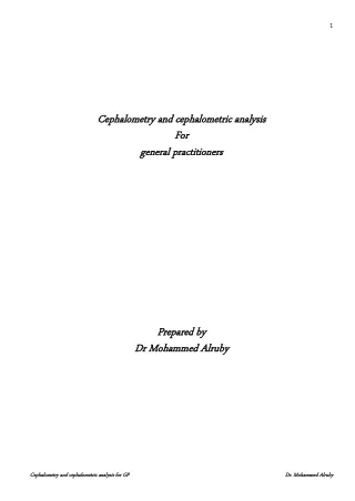

Cephalometry and Cephalometric analysis for General Practitioners
E N D
Presentation Transcript
1 Cephalometry and cephalometric analysis Cephalometry and cephalometric analysis For For general practitioners general practitioners Prepared by Prepared by Dr M Dr Mohammed ohammed Alruby Alruby Cephalometry and cephalometric analysis for GP Cephalometry and cephalometric analysis for GP Dr. Mohammed Dr. Mohammed Alruby Alruby
2 The assessment of Cranio- facial structures forms a part of orthodontic diagnosis. The discovery of X-rays in 1895 by Roentgen revolutionized dentistry. It provided a method of obtaining the inner Cranio – facial measurements with quite a bite of accuracy and reproducibility. In 1922 Paccini standardized the radiographic head images by positioning the subjects against a film cassette at a distance of 2 meters from the X-ray tube. In 1931 Broadbent in USA and Hofrath in Germany simultaneously presented a standardized cephalometric technique using a high powered X-ray machine and head holder called cephalostate. The term cephalometric is used to describe the analysis and measurements made on the cephalometric radiographs. Cephalogram: standardized radiograph of the head and face Standardization: = presence of head orientation for all subjects and for the same subject in the serial studies. =The target film distance was 60 inches= 5 feet = 180 cm. =from film to midsagittal plane= 15 cm. = the exposure time varies according to the age of the patient and usually from 1/2 to 3/4 second. Important of standardization: 1-Make it possible to study facial growth by taking a serial radiograph in a standard manner, thus any changes incorporated by growth can be detected. 2-Make it possible to localize the disease and the site of dentofacial deformities. 3-Comparisons of cephalogram before and after treatment thus the changes due to treatment can be detected. Uses of cephalometric in orthodontics: 1-Classification of dental and skeletal abnormalities. 2-growth studies. 3-Aids in treatment planning. 4-Evaluation of effectiveness of various orthodontic procedures. 5-Evaluation of effectiveness of retention. 6-Evaluation of growth changes after treatment was completed. Limitation, disadvantage of cephalometric: 1-It is two dimensional representations for three dimensional structures. 2-Superimpostion. 3-Degree of reliability of landmark as measuring points is still uncertain. 4-Locate the site of discrepancy but do not reveal the basic etiologic factors. 5-Magnification, Distortion and Blurring. Magnification: Proportional enlargement of all parts of structure in the Cephalometry. This error occurs because the X-ray beams are not parallel with all points of the object. We can minimize this error by using a long focus- object distance and a short film – object distance and by use of angular rather than linear measurements. Distortion: Lack of exact reproduction of a structure in the term of proportion. Magnification occurs when all parts of structure are increase proportionally, while in distortion, the different parts of structure are not increase proportionally. In lateral film, the only structure that not distorted are those Cephalometry and cephalometric analysis for GP Cephalometry and cephalometric analysis for GP Dr. Mohammed Dr. Mohammed Alruby Alruby
3 situated on the midsagittal plane (midline structure) while, all other bilateral structure is distorted. In anteroposterior films all structures are distorted. Blurring: Lack of sharpness of the radiographic image. Factors affecting blurring: 1-The movement of patient during exposure increase blurring. The patient must be instructing to close his teeth together into centric occlusion and do not move. 2-Sorce-object-film distance: the closer the object to the source, the greater the blurring, conversely the closer the object to the film, the lesser the blurring. 3-Time of exposure: decrease the time of exposure decrease the blurring. 4-The focal spot of radiation: the small the focal spot of radiation, the lesser the blurring. 5-Secondary radiation: the lesser secondary radiation, the lesser the blurring. Cephalometric landmarks: The cephalometric landmarks are of two types: 1-anatomical landmarks: these landmarks represent actual anatomic structures of the skull. 2-Derived landmarks: these are landmarks that have been obtained secondarily from anatomic structures in a cephalogram. The landmarks that are used in cephalometric should fulfil certain requirements: A-It should be easily seen in radiograph. B-It should be uniform in outline and should be reproducible. C-The landmarks should permit valid quantitative measurements of line and angles projected from them. The following are some of the important cephalometric landmarks: Nasion(N): The most anterior point midway between the frontal and nasal bones on the fronto - nasal suture. Orbitale(O): The lowest point on the inferior bony margin of the orbit. Porion (P): The most superior point on the external auditory meatus. Sella (S): The point representing the midpoint of the pituitary fossa or sella turcica. It is a constructed point in the mid-sagittal plane. Point A: it is the deepest point in the midline between the anterior nasal spine and alveolar crest between the two centeral incisors. Point B: It is the deepest point in the midline between the alveolar crest of the mandible and the mental process. Gonion (Go): The most everted point on the angle of the mandible formed by the junction between the ramus and the body of the mandible. Pogonion (Pog): The most anterior point on the symphysis of the mandible. Anterior nasal spine (ANS): The most anterior projection of the floor of the nasal cavity. Posterior nasal spine (PNS): The most posterior projection of the palatine bones in the midline of the roof of the oral cavity. Menton(Me): the most inferior midline point on the mandibular symphysis. Gnathion(Gn): It is the most anterior – inferior point on the symphysis of the chin. It is a constructed by intersecting a line drawn perpendicular to the line connecting menton and pogonion. Cephalometry and cephalometric analysis for GP Cephalometry and cephalometric analysis for GP Dr. Mohammed Dr. Mohammed Alruby Alruby
4 Articulare(Ar): It is a point at the junction of the posterior border of the ramus and the inferior border of the basilar part of the occipital bone. Condylion(Cd): the most superior point on the head of the condyle. Basion(Ba):It is the median point of the anterior margin of the foramen magnum. Bolton point (Bo): The highest point at the posterior condylar notch of the occipital bone. Pterygomaxillary fissure (Ptm): The contour of the fissure projected onto the palatal plane. The anterior wall represents the maxillary tuberosity outline, the posterior wall represents the anterior curve of the pterygoid process. This point corresponds to PNS. Incisor superius: Incisal tip of the crown of the most anterior maxillary central incisors. Incisor inferius: Incisal tip of the crown of the most anterior mandibular central incisors. Keyridge (KR): The most lower point on the contour of the shadow of the anterior wall of the infera-temporal fossa. Clinoidal (Cl): The most superior point on the contour of the anterior clinoid. Rhinion (Rh): The most anterior intersection of the nasal bones, which forms the tip of the bony nose. Soft tissue landmarks: Soft tissue nasion (N \): The most concave or retruded point in the tissue overlying the area of the fronto-nasal suture, the intersection of the Sn line with the soft tissue anterior to nasion. Nasal crown (Nc): A point along the bridge of the nose half away between soft tissue nasion and pronasale. Pronasal (Pn): The most prominent or anterior point of the nose. Subnasal (Sn): The point at which the nasal septum between the nostrils merges with the upper cutenous lip in the midsagittal plane. Soft tissue subspinale (A|): The point of greatest concavity in the midline of the upper lip between sub nasal and labrale superius. Labrale superius (LS): The most anterior point on the margin of the upper membranous lip. Stomion (St): The median point of the oral embrasure when the lips are closed. Labrale inferius (LI): The most anterior point on the margin of the lower membranous lip. Soft tissue submentale (B\): The point of the greatest concavity in the midline of the lower lip between the soft tissue chin and labrale inferius. Soft tissue pogonion(pog|):The most prominent or anterior point on the soft tissue chin in the midsagittal plane. Soft tissue Gnathion (Gn|): The midpoint between the most anterior and inferior points of the soft tissue chin in the midsagittal plane. Reference Lines and planes: Once the landmarks or "alphabet" of the cephalometric language is learned, these points are then connected to form the various lines and planes that are used in cephalometric. S – N line: The cranial base line between the center of sella turcica (S point) and the anterior point of the fronto-nasal suture (N). This represents the anterior cranial base. Bolton plane: this is a plane since it connects three points in space, the two Bolton points posterior to the occipital condyles and nasion. This is a representation of the cranial base which divides the cranium and facial structure. Frankfort horizontal plane (FH): this plane connects the lowest points of the orbits (orbital) and the superior points of the external auditory meatus (porion). Cephalometry and cephalometric analysis for GP Cephalometry and cephalometric analysis for GP Dr. Mohammed Dr. Mohammed Alruby Alruby
5 Palatal plane: although it only connects two points, the palatal "line" is often referred to as the palatal "plane". Nevertheless, it is important reference linking the anterior nasal spine (ANS) of the maxilla and the posterior nasal spine (PNS) of the palatine bone. Occlusal plane: this denture plane bisects the posterior occlusion of the permanent molars and premolars (or deciduous molars in mixed dentition) and extends anteriorly. In an ideal situation, the occlusal plane also bisects the occlusion of the incisor teeth. Mandibular plane: several mandibular planes are used, depending on the analysis. The most common ones are: a tangent to the lower border of the mandible; a line between gonion (GO) and gnathion (GN); or a line between gonion and menton (Me). It is usually not critical which is used, as long as the clinician uses the same one consistently in order not to incorporate error in a longitudinal study. Basion Nasion line: a line from basion to nasion representing the cranial base. Facial plane: a line from the anterior point of the frontonasal suture (N) to the most anterior point of the mandible (Pog). E line (E): a line between the most anterior points of the soft tissue of the nose and soft tissue of the chin. Cephalometric measurements: SNA: (average: 82 -+ 4). This measurement gives an indication of the anteroposterior position of apical base of maxilla to the cranial base line. This angle is larger than normal in case of Class II malocclusion. The angle is smaller than normal in Skeletal Class III malocclusion. SNB: (average: 79-+4) This measurement gives an indication of the antro-posterior position of apical base of the mandible to the cranial base line (SN). This angle is larger than normal in true Class III malocclusion. This angle is smaller than normal in Class II malocclusion. ANB: (average: 3-+1) This measurement reveals the maxillo mandibular relationship of upper and lowers apical bases. Larger angle than normal gives an indication to Class II malocclusion. Negative angle is indicating of sever Class III malocclusion but zero angle is indicates that malocclusion is more likely to be Class III. SNpog: (average 80-+4) This measurement gives an indication of the anteroposterior position of the mandible to the cranial base line (SN). The angle is larger than normal in true Class III malocclusion. The angle is smaller than normal in Class II malocclusion. Cephalometry and cephalometric analysis for GP Cephalometry and cephalometric analysis for GP Dr. Mohammed Dr. Mohammed Alruby Alruby
6 (Reference cephalometric points) (Cephalometric soft tissue points) (Cephalometric lines and planes) Cephalometry and cephalometric analysis for GP Cephalometry and cephalometric analysis for GP Dr. Mohammed Dr. Mohammed Alruby Alruby
7 Facial angle: Npog to FH plane (average: 87-+6) This angle gives an indication of the anteroposterior position of the most anterior point of the mandible to the upper face. Larger angle indicates of Class III malocclusion. Smaller angle indicates of Class II malocclusion. Gonial angle ar-Go-Me: (average 124 -+5) This angle gives an expression to the form of the mandible and plays a role in growth prognosis. Larger angle indicates more tendency to posterior relation of the mandible but smaller angle indicate more tendency to anterior relation of the mandible. SN to palatal plane: (average 9-+3) This angle gives an indication of the maxilla to the anterior cranial base. Larger angle indicate that the inclination is in anterior direction, but smaller angle indicate that the inclination is in posterior direction. Sn to mandibular plane: (average32-+3) This measurement gives an indication to the inclination of the mandible to the anterior cranial base. Larger angle indicate that the inclination is in posterior direction, but smaller angle indicate that the inclination is in anterior direction. Palatal plane to mandibular plane: (average 23-+4) This angle gives the relation between the maxillary and mandibular base. Larger angle indicate that there is an increase in lower anterior facial height, but smaller angle indicate that there is decrease in lower anterior facial height. FH plane to mandibular plane: (average: 25-+3) This angle gives the relation between the mandible and the upper face. Larger angle gives indication to the posterior inclination of the mandible; smaller angle gives indication to the anterior inclination of the mandible. This angle gives an idea about the vertical dysplasia. As the angle increased -------------------- open bite As the angle decreased --------------------- deep bite. Y axis to FH plane: (average 61-+4) This angle formed by the intersection between S- Gn and FH plane (lower anterior angle). This angle indicates the downward and forward position of the chin in relation to upper face at FH plane, it indicates the mandibular growth direction. Upper incisors to FH plane: (average 112-+6) This angle reveals the inclination degree of the central incisors as related to FH plane. Larger angle is usually a characteristic of skeletal Class II division 1 and Class III malocclusion. Smaller angle is indicative in cases of Class II division 2 malocclusion. Upper incisors to palatal plane: (average 109-+4) This angle reveals the inclination degree of central incisors as related to palatal plane. Larger angle ------------Class II division 1 malocclusion -------------- Class III malocclusion Smaller angle ------------Class II division 2 malocclusion Upper incisors to SN plane: (average 104 -+4) Cephalometry and cephalometric analysis for GP Cephalometry and cephalometric analysis for GP Dr. Mohammed Dr. Mohammed Alruby Alruby
8 This angle reveals the inclination degree of the centeral incisors as related to the cranial base line (SN). Larger angle ------------------ Class II division 1 -------------------- Class III malocclusion Smaller angle------------------ Class II division 2 malocclusions. Upper incisors to NA line: (average 25_+6) This angle indicates the degree of inclination of upper incisors in relation to NA line. Larger angle seen in Class II division 1 malocclusion. Smaller angle indicates retrusion of upper incisors. Lower incisors to mandibular plane: (average 95 -+5) Angle formed between the long axis of lower centeral incisors and the mandibular plane. Larger angle indicates a protrusive direction as in case of Class I bimaxillary protrusion. Smaller angle indicates a retrusive direction as in case of Class III malocclusion. Lower incisors to FH plane: (average 59-+4) Angle formed between the long axis of lower central incisors to FH plane. This angle reveals the relation between lower incisors and upper face, larger angle indicates more retrusive position of lower incisors but smaller angle indicates more protrusive position of lower incisors. Inter incisal angle: (average 127 _+8) Angle formed between the long axis of lower and upper incisors. Larger angle reveals more upright position of the incisors as in case of Class II division 2 malocclusion. Smaller angle reveals more protruded incisors as in case of bimaxillary protrusion. Lower incisors to NB line: (average: 29_+6) This angle reveals the inclination degree between the long axis of lower incisors and NB line. Larger angle seen in Class II division 1 malocclusion. Smaller angle seen in Class III malocclusion. Anterior facial height: Total anterior facial height: from nasion to menton Upper anterior facial height: from nasion to ANS and this equal 45% from total. Lower anterior facial height: from ANS to menton and this equal 55% from total. Posterior facial height: from S point to gonion and equal 65 % from total anterior facial height. Molar relation: the distance between the distal surfaces of the upper and lower molars measured along the occlusal plane. This measurement gives an indication of the extent of the horizontal malocclusion. The norm (-3.0mm -+3.0mm). Incisor over jet: the distance between the incisal tips of the upper and lower incisors measured along the facial plane and measures the horizontal dimension. Incisor over bite: the distance between the upper and lower incisors tip along the facial plane and measure the vertical distances. Soft tissue analysis: Cephalometry and cephalometric analysis for GP Cephalometry and cephalometric analysis for GP Dr. Mohammed Dr. Mohammed Alruby Alruby
9 Soft tissue convexity: (N/ Prn /Pog) (average=135): This measurement gives an indication of the convexity or concavity of the soft tissue profile with the nose included. Because this measurement is directly affected by mandibular growth, retrognathism (Class II division 1) is associated with convex soft tissue profile. Length of upper lip: stomion to subnasal, average (24mm in male and 20mm in females) Length of lower lip: stomion to gnathion, average (50mm in male and 46 in females). E line and lips: lips to Prn Pog line, average (upper lip= 1mm behind; lower lip= 0mm) These measurements indicate the anteroposterior position of the lips with reference to a line between the most anterior part of soft tissue chin (pogonion) and the most anterior part of the nose (pronasal). Dentures that are forward (Class I bimaxillary protrusion and Class II division 1 malocclusion) produce a convex profile with the lips a head of the E line. Straight or concave profiles (Class II division 2 or Class III malocclusion) are associated with retruded lips. Nasolabial angle: the angle formed by tangent to the lower border of the nose and tangent to the upper lip. The average norm (90- 110) degree. Equipment and material for tracing: View box, preferably variable light intensity. 0.003- inch thick tracing acetate with one matte surface. Millimeter ruler. Protractor. Compass. Draftsman triangle. Well- sharpened medium (no.0.3 pencil) The tracing acetate is customarily attached to the film with two small pieces of masking tape along on edge. Cephalometry and cephalometric analysis for GP Cephalometry and cephalometric analysis for GP Dr. Mohammed Dr. Mohammed Alruby Alruby
10 (SNA angle) (SNB angle) (ANB angle) (Upper incisor to NA line) Cephalometry and cephalometric analysis for GP Cephalometry and cephalometric analysis for GP Dr. Mohammed Dr. Mohammed Alruby Alruby
11 (Y axis angle (SGn to FH) Posterior to anterior facial height (S go to N Me) Cephalometry and cephalometric analysis for GP Cephalometry and cephalometric analysis for GP Dr. Mohammed Dr. Mohammed Alruby Alruby
12 (Tweed triangle) (Sn to go Gn) (Relation between upper and lower lip to E line in case of bimaxillary protrusion) Cephalometry and cephalometric analysis for GP Cephalometry and cephalometric analysis for GP Dr. Mohammed Dr. Mohammed Alruby Alruby
13 (Length of upper and lower lip) \ (Nasolabial angle) (Molar relation) Cephalometry and cephalometric analysis for GP Cephalometry and cephalometric analysis for GP Dr. Mohammed Dr. Mohammed Alruby Alruby
14 (Upper and lower gonial angle) (Facial angle) Cephalometry and cephalometric analysis for GP Cephalometry and cephalometric analysis for GP Dr. Mohammed Dr. Mohammed Alruby Alruby