Download
1 / 5
50 likes | 65 Views
Direct Plagiarism

E N D
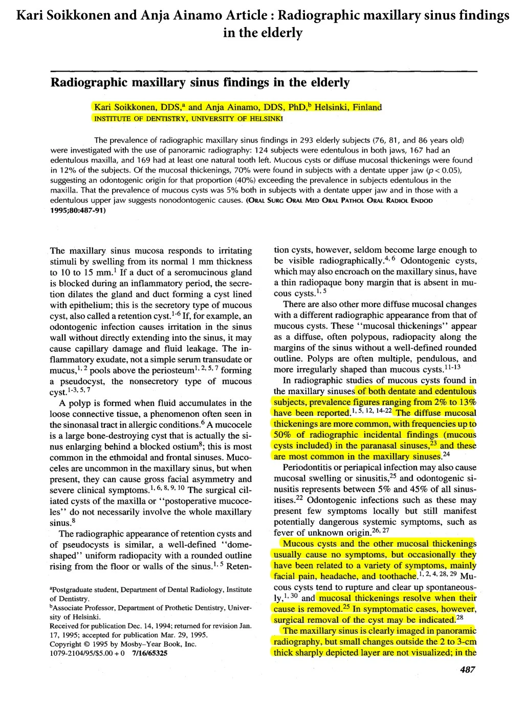
Kari Soikkonen and Anja Ainamo Article : Radiographic maxillary sinus findings in the elderly Radiographic maxillary sinus findings in the elderly Kari Soikkonen, DDS, a and Anja Ainamo, DDS, PhD, b Helsinki, Finland INSTITUTE OF DENTISTRY, UNIVERSITY OF HELSINKI The prevalence of radiographic maxillary sinus findings in 293 elderly subjects (76, 81, and 86 years old) were investigated with the use of panoramic radiography: 124 subjects were edentulous in both jaws, 167 had an edentulous maxilla, and 169 had at least one natural tooth left. Mucous cysts or diffuse mucosal thickenings were found in 12% of the subjects. Of the mucosal thickenings, 70% were found in subjects with a dentate upper jaw (p < 0.05), suggesting an odontogenic origin for that proportion (40%) exceeding the prevalence in subjects edentulous in the maxilla. That the prevalence of mucous cysts was 5% both in subjects with a dentate upper jaw and in those with a edentulous upper jaw suggests nonodontogenic causes. (ORAL SURG ORAL MED ORAL PATHOL ORAL RADIOL ENDOD 1995;80:487-91 ) tion cysts, however, seldom become large enough to be visible radiographically. 4,60dontogenic cysts, which may also encroach on the maxillary sinus, have a thin radiopaque bony margin that is absent in mu- cous cysts. 1' 5 There are also other more diffuse mucosal changes with a different radiographic appearance from that of mucous cysts. These "mucosal thickenings" appear as a diffuse, often polypous, radiopacity along the margins of the sinus without a well-defined rounded outline. Polyps are often multiple, pendulous, and more irregularly shaped than mucous cysts. 11-13 In radiographic studies of mucous cysts found in the maxillary sinuses of both dentate and edentulous subjects, prevalence figures ranging from 2% to 13% have been reported, l, 5, 12, 14-22 The diffuse mucosal thickenings are more common, with frequencies up to 50% of radiographic incidental findings (mucous cysts included) in the paranasal sinuses, 23 and these are most common in the maxillary sinuses. 24 Periodontitis or periapical infection may also cause mucosal swelling or sinusitis, 25 and odontogenic si- nusitis represents between 5% and 45% of all sinus- irises. 22 Odontogenic infections such as these may present few symptoms locally but still manifest potentially dangerous systemic symptoms, such as fever of unknown origin. 26, 27 Mucous cysts and the other mucosal thickenings usually cause no symptoms, but occasionally they have been related to a variety of symptoms, mainly facial pain, headache, and toothache. 1, 2, 4, 28, 29 MU- COUS cysts tend to rupture and clear up spontaneous- ly,1, 30 and mucosal thickenings resolve when their cause is removed. 25 In symptomatic cases, however, surgical removal of the cyst may be indicated. 28 The maxillary sinus is clearly imaged in panoramic radiography, but small changes outside the 2 to 3-cm thick sharply depicted layer are not visualized; in the The maxillary sinus mucosa responds to irritating stimuli by swelling from its normal 1 mm thickness to 10 to 15 mm. 1 If a duct of a seromucinous gland is blocked during an inflammatory period, the secre- tion dilates the gland and duct forming a cyst lined with epithelium; this is the secretory type of mucous cyst, also called a retention cyst. 1-6 If, for example, an odontogenic infection causes irritation in the sinus wall without directly extending into the sinus, it may cause capillary damage and fluid leakage. The in- flammatory exudate, not a simple serum transudate or mucus,l, 2 pools above the periosteum 1' 2, 5, 7 forming a pseudocyst, the nonsecretory type of mucous cyst. 1-3, 5, 7 A polyp is formed when fluid accumulates in the loose connective tissue, a phenomenon often seen in the sinonasal tract in allergic conditions. 6 A mucocele is a large bone-destroying cyst that is actually the si- nus enlarging behind a blocked ostium8; this is most common in the ethmoidal and frontal sinuses. Muco- celes are uncommon in the maxillary sinus, but when present, they can cause gross facial asymmetry and severe clinical symptoms. 1, 6, 8, 9, 10 The surgical cil- iated cysts of the maxilla or "postoperative mucoce- les" do not necessarily involve the whole maxillary sinus, s The radiographic appearance of retention cysts and of pseudocysts is similar, a well-defined "dome- shaped" uniform radiopacity with a rounded outline rising from the floor or walls of the sinus. 1' 5 Reten- apostgraduate student, Department of Dental Radiology, Institute of Dentistry. bAssociate Professor, Department of Prothetic Dentistry, Univer- sity of Helsinki. Received for publication Dec. 14, 1994; returned for revision Jan. 17, 1995; accepted for publication Mar. 29, 1995. Copyright ?9 1995 by Mosby-Year Book, Inc. 1079-2104/95/$5.00 + 0 7/16/65325 487
Kari Soikkonen and Anja Ainamo Article : Radiographic maxillary sinus findings in the elderly 488 ORAL SURGERY ORAL MEDICINE ORAL PATHOLOGY Soikkonen andAinamo October 1995 Table I, Subjects of the study Age in years 76 81 86 , Total Men Women Total 44 (15%) 106 (36%1 150 (51%) 28 (10%) 59 (20%) 87 (30%) 14 f5%~ 42 (14%) 56 (19%) 86 (29%) 207 (71%) 293 (100%) Table II. Maxillary sinus findings (N) according to age Age in years 76 81 86 Total Number Percentage Number Percentage Number Percentage Number Percentage Normal Mucous cyst Diffuse mucosal thickening Total 127 86 5 9 10"0 77 1 8 8-6 89 1 9 10~ 48 6 87 11 2 10--6 252 14 23 28"--9 87 5 8 l"-~ 7 14 14-8 1 5-5 In four cases sinuses were not diagnostically depicted. Table III, Maxillary sinus findings according to sex Women Men Total Number I Percentage Number Percentage Number Percentage Normal Mucous cyst Diffuse mucosal thickening Total 183 89 4 7 100 69 5 __9 83 83 6 l 1 100 252 14 23 289 87 5 __8 100 9 14_ 206 In four cases sinuses were not diagnostically depicted. normal panoramic projection, the roof of the maxil- lary sinus is not imaged. 22 However. mucous cysts and other mucosal thickenings are usually well dem- onstrated, as they almost always arise from the antral floor.i, 4. 13. 17.31.32 Few studies have thus far been made to assess the prevalence of maxillary sinus findings in the elderly. The aim of the present study was to investigate, with the use of panoramic radiography, the preva- lence of maxillary sinus findings in elderly subjects aged 76, 81. and 86 years, and to test the hypothesis that such findings are more prevalent in dentate sub- jects. was 81.8% (n = 651). 33 Between 1989 and 1990. 651 subjects participated in the medical examination. Be- fore May 31, 1990 the mortality among the partici- pants in the medical survey was 8.0% (n = 51). In 1990 and 1991, the 600 subjects who were still alive were invited to the Institute of Dentistry for a com- prehensive dental examination. A total of 133 of the 600 were interviewed only by phone or by mail, and no dental data were available for 103 subjects. Alto- gether 364 subjects remained. 28% were men and 72% women; they were examined in 1990 and 1991.34 The response rate for invited men was 69% and for women 58%. Of the 364 subjects, 293 attended the Institute of Dentistry and were examined clinically and radiographically, whereas 71 subjects were ex- amined clinically only in their homes or in institu- tions. The subjects were radiographed between the months of August 1990 and June 1991. Of the 293 subjects radiographed (86 men and 207 women) (Table I), 169 (54 men and 115 women) had one or more clinically visible natural teeth left. They MATERIAL AND METHODS This investigation is a part of a large Finnish med- ical and dental survey of a random sample of elderly subjects (total, 8035) who were born in 1904, 1909, and 1914 and living in Helsinki in January 1989. 33 The response rate for the medical survey from the random sample of 795 elderly invited to participate
Kari Soikkonen and Anja Ainamo Article : Radiographic maxillary sinus findings in the elderly 489 ORAL SURGERY ORAL MEDICINE ORAL PATHOLOGY Volume 80, Number 4 Soikkonen and Ainamo Table IV. Maxillary sinus findings in clinically edentulous or dentulous subjects Edentulous Dentulous Total Number Percentage Number I Percentage Number I Percentage Normal Mucous cyst Diffuse mucosal thickening Total 116 93 3 3 100 136 10 19 165 82 6 12 100 252 14 23 289 87 5 8 100 4 4 124 Table V. Maxillary sinus findings in subjects with edentulous or dentulous maxilla Edentulous maxilla Dentulous maxilla Total Number Number Percentage Percentage Number Percentage Normal Mucous cyst Diffuse mucosal thickening Total 150 91 5 4 100 102 82 5 13 100 252 14 23 289 87 5 8 100 8 7 6 16 124 165 Total chi-square: 7.28, p = 0.0262. formed the dentulous subgroup. The mean number of teeth in this group was 17.0 for men and 14.5 for women, 35 with 124 subjects (32 men and 92 women) clinically edentulous. 36 Patients with at least one ra- diographically visible natural tooth or root in the up- per jaw numbered 126 (44 men, 82 women), whereas 167 (42 men, 125 women) were radiographically edentulous in the upper jaw with not even any impacted teeth or retained roots in the maxilla. Panoramic radiographs were taken with a PM 2002 radiographic apparatus (Planmeca Oy, Helsinki, Fin- land). Trimax T16 intensifying screens and GTU x- ray film (3M Co., St. Paul, Minn.) were used. Intraoral radiographs of areas poorly visible in the panoramic radiograph were taken using a Siemens Heliodent 70 dental radiographic unit (Siemens Med- ical Engineering, Dental Sector, Bensheim, Ger- many) and Kodak Ultra-speed x-ray film (Eastman Kodak Co., Rochester, Minn.). In all, 169 panoramic and 109 intraoral radiographs were taken. All films were developed by automatic processing. Intraoral films were mounted in frames. The radiographs were studied by one dental radiologist (K.S.) under stan- dardized conditions with the use of Mattson' s binoc- ulars (X-Produkter, Maim6, Sweden) with a x2 mag- nification and a viewing light of adjustable brightness when necessary. Findings of increased radiopacity in the maxillary sinuses were recorded and divided into two catego- ries: mucous cysts, well-defined radiopacities with a rounded (convex) outline rising from the floor or walls of the sinus; and mucosal thickenings, which Table VI. Intra-examiner variation in re-examination of 41 patient radiographs Round 2 I Normal Finding Total Normal Finding Total 35 0 35 1 5 6 36 5 41 Round 1 K: 0.58, fair agreement. represented the more diffuse radiopacities along the margins of the sinus without well-defined rounded outlines. The intra-examiner variation was assessed by re-examining the radiographs of 41 randomly se- lected patients (21 dentulous, 20 edentulous), The in- terval between the two rounds of viewings was 6 months. The manually calculated Cohen's kappa test was used to determine the statistical significance of the intra-examiner agreement. The other data were ana- lyzed with the chi square test, and the StatView SE+ Graphics (Abacus Concepts Inc., Berkeley, Calif.) statistical program package for the Macintosh com- puter (Apple Computer Inc., Cuppertino, Calif.). RESULTS In the material as a whole, mucous cysts or mucosal thickenings were found in 37 subjects (12%). In 14 subjects (5%) the findings were classified as mucous cysts. Diffuse mucosal thickenings were found in 23
Kari Soikkonen and Anja Ainamo Article : Radiographic maxillary sinus findings in the elderly 490 ORAL SURGERY ORAL MEDICINE ORAL PATHOLOGY Soikkonen andAinamo October 1995 cous cysts showed no age-dependent tendencies. The diffuse mucosal thickenings, however, were slightly more prevalent in the younger age groups, who had more teeth. The majority of both the mucous cysts and the diffuse mucosal thickenings were found in dentate subjects. However, the same percentage of mucous cysts (5%) was observed in subjects with natural teeth in the upper jaw and in those with an edentulous up- per jaw. From the similar prevalences of mucous cysts in the two groups it can be suspected that odontogenic causes may not be a major contributing factor in their formation. The prevalence and size of mucous cysts in sites of periapical or periodontal pathosis and in sites without pathologic findings have also previously been found to be similar. 21 Neither that finding nor ours supports the findings of Halstead, 3~ who reported that a possible odontogenic cause could be indicated in 90% of subjects with mucous cysts. However. in the present study, 70% of the diffuse mucosal thick- enings were found in subjects who had at least one radiographically visible natural tooth or root in the upper jaw. Thus it may be suspected that the diffuse thickenings may be odontogenic in origin for that proportion (40%) of subjects exceeding the preva- lence of the same findings in the sinuses of subjects with no maxillary teeth at all. The presence of mucosal thickenings in the max- illary sinus floor always indicates the presence of ir- ritative stimuli, often an infection of dental origin. Such infective foci (chronic apical periodontitis, deep infrabony pockets caused by periodontitis) are usu- ally unaccompanied by any major subjective symp- toms. Their accurate diagnosis may sometimes be vi- tal to the patient, for if the host resistance decreases for some reason, it will give these infections an op- portunity to become exacerbated and cause acute si- nusitis, whereas the possibility also exists of further spread and systemic manifestations. 26 27 cases (8%). The sex and age distributions of the ra- diographic maxillary sinus findings are summarized in Tables II and HI. Seventy-one percent of the mu- cous cysts and 83% of the diffuse mucosal thicken- ings were found in clinically dentate subjects (Table iv). Twenty-two of the subjects with at least one radio- graphically visible natural tooth or root in the upper jaw had radiopaque findings in their maxillary sinuses (18%). In six of these subjects (5%), the findings were classified as mucous cysts. Diffuse mucosal thicken- ings were found in 16 cases (13%) (Table V). Seventy percent of the diffuse mucosal thickenings were found in subjects with at least one radiographically visible natural tooth or root in the upper jaw (p < 0.05). The prevalence of mucous cysts was 5% in both sub- jects with at least one radiographically visible natural tooth or root in the upper jaw and those with completely edentulous upper jaw. No statistically significant differences were found between the sexes or between age groups, although the prevalence of diffuse mucosal thickenings dimin- ished slightly with age. No destructive mucoceles were found. In four cases the sinuses were not diag- nostically depicted. The intra-examiner agreement between the two rounds of viewings was fair, (K = 0.58) (Table VI). DISCUSSION The prevalence of mucous cysts and diffuse mu- cosal thickenings in the maxillary sinuses of our eld- erly edentulous subjects was 7%. However, figures ranging from 2.6% to 20% have been reported from other studies of edentulous subjects. 37" 38 Studies of rounded shadows (mucous cysts) in maxillary sinuses found, in both dentate and edentulous subjects, figures ranging from 2% to 13%.1.5. 12. 14-22 Our figure of 5% for the prevalence of mucous cysts falls within that range. The preva- lence of mucous cysts and diffuse mucosal thicken- ings in all the paranasal sinuses together has occa- sionally been as high as 50% in facial radiographs taken for indications other than suspected sinus dis- ease. 23 In a magnetic resonance imaging study of in- cidental findings in the paranasal sinuses of 438 sub- jects, the prevalence of incidental findings in all sinuses was 37.5%; and they were most common in the maxillary sinuses (27%). 24 According to Mattila 17 the prevalence of mucous cysts is not age-dependent. In studies including younger age groups, mucous cysts have been most prevalent in the third decade, and they have also been found to be more prevalent in men.X, 5, 22 In the rather narrow age-range of the present study's very old subjects, the number of mu- REFERENCES 1. Goaz P, White S. Oral radiology: principles and interpreta- tion. 3rd ed. St Louis: CV Mosby, 1994:602-10. 2. Lindsay JR. Nonsecreting cysts of the maxillary sinus mucosa. Laryngoscope 1942:52:84-100. 3. Schuknecht HF. Lindsay JR. Benign cysts of the paranasal sinuses. Arch Otolaryngol 1949;49:609-30. 4. Paparella MM. Mucosal cyst of the maxillary sinus: diagno- sis and management. Arch Otolaryngol 1963;77:650-7. 5. Allard RHB, van der Kwast WAM, van der Waal I. Mucosal antral cysts: review of the literature and report of a radio- graphic survey. OgAL SURG ORAL MED ORAL PATHOL 1981; 51:2-9. 6. Gardner DG. Pseudocysts and retention cysts of the maxillary sinus. ORAL SURG ORAL MED ORAL PATHOL 1984;58:561-7. 7. McGregor GW. Formation and histologic structure of cysts of the maxillary sinus. Arch Otolarygol 1928;8:505-19.
Kari Soikkonen and Anja Ainamo Article : Radiographic maxillary sinus findings in the elderly Soikkonen and Ainamo 491 ORAL SURGERY ORAL MEDICINE ORAL PATHOLOGY Volume 80, Number 4 8. Gardner DG, Gullane PJ. Mucoceles of the maxillary sinus. OPAL SURG ORAL MED ORAL PATHOL 1986;62:538-43. 9. Ormerod LD, Weber AL, Ranch SD, Feldon SE. Ophtalmic manifestations of maxillary sinus mucoceles. Ophtalmology 1987;94:1013-9. 10. Salam MA, Whitehead E. Large antral maxillary mucocele presenting with facial asymmetry. J Laryngol Otol 1993; 107:451-2. 11. Killey HC, Kay LW. Benign mucosal cysts of the maxillary sinus. Int Surg 1970;53:235-44. 12. Wright RW~ Round shadows in the maxillary sinuses. Laryn- goscope 1946;56:455-89. 13. Kwapis B J, Whitten JB. Mucosal cysts of the maxillary sinus. J Oral Surg 1971;29:561-6. 14. Ibsen B. Basal convex shadows in maxillary sinus. Nord Med 1945;27:1487-9. 15. Kivim~iki J, Ekholm A. Klinisch-pantomographische Unter- suchungen uber Schleimhauteretentionzysten der Kieferh61e. Proc Finn Dent Soc 1961;57:3-13. 16. Myrhaug H. Behandling av odontogene affeksjoner av kjeve- huten. Norske Tannlaegeforen Tid 1962;72:211-8. 17. Mattila K. Roentgenological investigations into the relation between periapical lesions and conditions of the mucous membrane of maxillary sinuses. Acta Odont Scand 1965;23 (suppl 42): 1-91. 18. Lilly GE, Cutcher JL, Steiner M. Spherical shadows within the maxillary antrum. J Oral Med 1967;23:19-21. 19. Myall RWT, Eastep PB, Silver JG. Mucous retention cyst of the antrum. J Am Dent Assoc 1974;89:1338-42. 20. Ohba T. Manson-Hing LR. Radiological study of cyst-like lesions in the maxillary sinus. Dentomaxillofac Radiol 1975; 4:100-3. 21. MacD~176 DS" Muc~ population. Dentomaxillofac Radiol 1993;22:208-10. 22. McGowan D, Baxter P, James J. The maxillary sinus and its dental implications. Oxford:Wright, 1993:1-153. 23. Wilson PS, Grocutt M. Mucosal thickening on sinus X-rays and its significance. J Laryngol Otol 1990;104:694-5. ?9 24. Cooke LD, Hadley DM. MRI of the paranasal sinuses: inci- dental abnormalities and their relationship to symptoms. J Laryngol Otol 1991;105:278-81. 25. Falk H, Ericson S, Hugoson A. The effects of periodontal treatment on mucous membrane thickening in the maxillary sinus. J Clin Periodontol 1986;13:217-22. 26. Naschitz JE, Yeshurun D. Occult infection in the facial area presenting as fever of unknown origin. Isr J Med Sci 1985; 21:995-8. 27. Huebner GR, Groat D. The role of dental disease in fever of unknown origin. Postrgrad Med 1986;79:275-8. 28. Fisher EW, Whittet HB, Croft CB. Symptomatic mucosal cysts of the maxillary sinus: antroscopic treatment. J Laryn- gol Otol 1989;103:1184-6. 29. Rhodus NL. The prevalence and clinical significance of max- illary sinus mucous retention cysts in a general clinic popu- lation. Ear Nose Throat J 1990;69:82-7. 30. Halstead CL. Mucosal cysts of the maxillary sinus: report of 75 cases. J Am Dent Assoc 1973;87:1435-41. 31. Ohba T, Katayama H. Comparison of panoramic radiogra- phy and Water's projection in the diagnosis of maxillary sinus disease. ORAL SURG ORAL MED ORAL PATHOL 1976;42: 534-8. 32. Ohba T. Value and limitation of panoramic radiography in the diagnosis of maxillary sinus pathosis. Int J Oral Surg 1977; 6:211-4. 33. Valvanne J. The prognostic significance of clinical findings in the elderly: a one-year follow up study of groups of people aged 75, 80 and 85 years living in Helsinki. Helsinki, Finland; University of Helsinki: 1992: Thesis. 34. Ainamo A, Hiltunen K, Nevalainen J, et al. Helsinkil~iisten vanhusten suunterveys. Alustava raportti. Suomen Hammas- laakarilehti 1993;40:1176-92. 35. Ainamo A, Soikkonen K, Wolf J, et al. Dental radiographic findings in the elderly in Helsinki, Finland. Acta Odontol Scand 1994;52:243-9. 36. Soikkonen K, Ainamo A, Wolf J, et al. Radiographic findings in the jaws of clinically edentulous old people living at home in Helsinki, Finland. Acta Odontol Scand 1994;52:229-33. 37. Tronje G, Bolin A, Eliasson S, Julin P. Panoramic radiogra- phy of edentulous jaws: I. frequency and distribution of pathological findings. Dentomaxillofac Radiol 1980;9:21-5. 38. Keur JJ, Campbell JPS, McCarthy JF, Ralph WJ. Radiolog- ical findings in 1135 edentulous patients. J Oral Rehabil 1987; 14:183-91. antral cysts in a Chinese Reprint requests: Kari Soikkonen, DDS Institute of Dentistry Department of Dental Radiology P.O. Box 41 (Mannerheimintie 172) FIN-00014 University of Helsinki Finland