Hebbian Plasticity
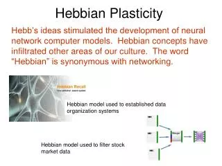
Hebb’s ideas stimulated the development of neural network computer models. Hebbian concepts have infiltrated other areas of our culture. The word “Hebbian” is synonymous with networking. Hebbian Plasticity. Hebbian model used to established data organization systems.

Hebbian Plasticity
E N D
Presentation Transcript
Hebb’s ideas stimulated the development of neural network computer models. Hebbian concepts have infiltrated other areas of our culture. The word “Hebbian” is synonymous with networking. Hebbian Plasticity Hebbian model used to established data organization systems Hebbian model used to filter stock market data
Who is Donald Hebb? • Student of Karl Lashley, the father of the Engram • Hebb worked with Penfield at McGill University and Montreal Neurological Institute and later was the Chairperson of the Psychology Department at McGill. • Author of Organization of Behavior in 1949 and the founder of the field of Biopsychology • His work is now recognized as seminal. So, what did he do?
His message about the Engram was twofold: • The engram could be widely distributed among connections that link the cells in the assembly of cells in the brain (see next slide) 2. The engram could involve the same neurons that are involved in sensation & perception (refer to the infereotemporal slide of the car and bird experts). So, destruction of only a fraction of the cells would not be expected to eliminate the memory, which is also what Lashley found even though his principles were not completely on target (i.e. all cortical areas do not contribute equally to memory – as you know, only the ones involved in the learning). Remember, Lashley had trouble destroying the “memory” in the rat after learning because the rats were learning the maze by sight, feel and smell (that is, many neural networks were involved), so damage to one was compensated by another system. How does the neural networking work….?
First, the Cell Assembly Neurons that are related functionally are linked neurally. This could be the primary visual association cortex (striate cortex) with the inferotemporal cortex and the subsequent activation of the hippocampus.
From Activation to Cell Strengthening The stimulus is the circle, which activates the cells. Cells continue to fire even after the stimulus is removed. Cells active at the same time will be strengthened. This is where memories are located, that is, within the “Hebbosomes” or protein molecules that are formed at the same time. No External Stimulus
Activation after Learning After learning all you need is stimulation of part of the neural network to retrieve the memory.
Associative vs. NonAssociative Learning Associative Learning (conditioning) is different from NonAssociative Learning (habituation and sensitization). Associative Learning involves the simultaneous or close occurring activation of two synapses in either the form of Classical Conditioning (Aplysia model) or Instrumental Conditioning. NonAssociative Learning is a very basic learned response, and has been used as model to understand what happens at the level of the synapse during simple learning.
Two Types of Associative Learning(just a refresher) Classical Conditioning: Condition (UCS) BEHAVIOR (gill withdrawal) Condition 2 (becomes CS) (touching siphon skin) Condition 2 can be given on it’s own once the association has been made, that is, the neural changes have taken place Instrumental Learning: Condition BEHAVIOR Consequence Reinforcement The outcome or the consequence increases or decreases the likelihood that the condition will elicit the same behavior (response) again.
In Associative learning (classical conditioning) there is a strong stimulus (US) that is paired with a weak stimulus (CS) General Model Proposed by Hebb Model depicted in Classical Conditioning terms from LeDoux, 2002 How does cell A “remember” what it has learned?
In this diagram consider cell A from the last slide to be the neuron in the center. Strong stimulus creates a tag at the synapse and initiates the synthesis of the protein molecule. Weak stimulus creates the tag only. When the strong stimulus and the weak stimulus are paired, the tag at the weak site can now utilize the protein (hence associative learning). From: LeDoux page 152
Aplysia (snail) used as a model for Classical Conditioning siphon Sunglasses not usually found on the average snail tail Diagram on the right shows the external anatomy. The gill will retract when water is sprayed on the skin the same way you will blink if an air puff strikes your eye. Very mild stimulation will result in indiscernible movement of the gill.
Aplysia: Neural Organization The sensory neuron is circled. Notice that it synapses (releases GLU) on a motor neuron that connects with the muscle that controls the gill response. The gill is sensitive to touch (e.g. spraying of water), which makes the gill retract, but light touch has no affect.
Aplysia: Neural Organization Another neuron circled in blue contains serotonin (L29 neuron) and is connected to the snail’s tail. A strong stimulus to the tail (e.g., shock) will causes changes in the sensory neuron via the axoaxonic synapse.
Here are the important players: CS (light touch to the siphon – does not elicit gill withdrawal response) US (shock to the tail – elicits a mild gill withdrawal response via it’s effect on the sensory neuron) L29 (also called facilitatory interneuron by LeDoux) releases Serotonin to terminal of the sensory neuron. Sensory Neuron releases Glutamate onto the Motor Neuron. To keep the diagram on the right consistent with LeDoux’s diagram on page 164, notice that a tail skin sensory neuron has been drawn. You will not find this on the other diagrams. US siphon Tail shock Light touch Sensory neuron interneuron
The shock to the tail causes L29 (interneuron) to release Serotonin on to the terminal of the sensory neuron. The activation of the G-protein on the Serotonergic metabotropic receptor activates Adenylyl cyclase, which in turn activates Protein Kinase. The Protein Kinase alters the K+ channel (adds a phosphate group), causing it to close. By keeping the K+ in the terminal of the sensory neuron, the cell fires longer. Ca++ channels then remain open because the charge is elevated and more Glu is released on to the motor neuron and the gill retracts. L29 terminal Sensory neuron terminal (releases GLU on to the motor neuron)
The CS is a very light touch to the sensory neuron that elicits no response on its own. Pairing the US and CS causes the gill to retract in an exaggerated manner. This is the same as the last slide. The shock to the tail is the US and this causes changes in the sensory neuron. L 29 neuron Sensory Neuron terminal
If the CS (light touch) is given alone (black) there is no response. If the US (shock to the tail) is given alone (red) there is a mild response. When the US and CS are paired the response is exaggerated. After training, the CS alone (light touch) that failed to elicit a response now elicits a strong response (not shown). US + CS US alone CS alone
Light Removal of the US results in the CS (light touch) causing the gill to retract. The means that synaptic changes have taken place that involve structural changes. Eric Kandel who pioneered this research beginning 40 years ago was awarded the Nobel Prize in Medicine in 2004 because of the significance of his work (it took that long for the importance to be recognized). In mammalian systems, we know that theses structural changes are in the form of alterations in protein synthesis.
The mammalian system involves insertion of more AMPA receptors and/or increasing sensitivity of the AMPA receptors. In order for this to happen, a number of molecular changes have to occur to induce the transcription of proteins that ultimately make the receptors and other permanent membrane changes that “hold our memories.”
Neural Changes in Memory are Accompanied by Molecular Changes • “persistently activated” Protein Kinases • Activation of CREB (that is, Cyclic-AMP Response Element Binding protein) The end result: Increase in Protein Synthesis and lifetime (rather than transient) structural changes that are thought to form our memories. (see ANGELL for two brief readings from Bear et al., 2007)