Download

1 / 3
0 likes | 1 Views
Advances in imaging technology have transformed the landscape of pituitary adenoma treatment. From early detection to precise surgical planning and long-term monitoring, imaging serves as the foundation of modern patient care.

E N D
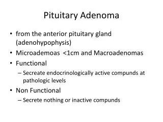
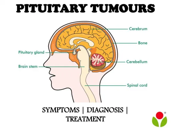
Advances in Imaging for Pituitary Adenoma Diagnosis and Treatment Pituitary adenomas are among the most common tumors of the brain, often arising from the pituitary gland at the base of the skull. While many of these tumors are benign, their presence can cause significant health issues due to hormonal imbalances and pressure on surrounding structures such as the optic nerves. With the evolution of modern imaging techniques, the ability to diagnose and guide pituitary adenoma treatment has advanced dramatically, offering patients improved outcomes and personalized care. Understanding Pituitary Adenomas What Are Pituitary Adenomas? Pituitary adenomas are tumors that originate in the pituitary gland. They can be classified as functional or nonfunctional, depending on whether they produce excess hormones. Functional adenomas may result in conditions such as Cushing’s disease or acromegaly, while nonfunctional ones can grow silently until they cause vision problems or headaches. 1/3
Why Imaging Matters Accurate imaging plays a central role in pituitary adenoma treatment. It helps doctors not only detect the tumor but also evaluate its size, location, and impact on surrounding brain structures. Imaging is also critical for surgical planning and monitoring after treatment. Advances in Imaging Technology Magnetic Resonance Imaging (MRI) MRI remains the gold standard in pituitary imaging. Advances in high-resolution MRI provide clearer visualization of microadenomas that were once difficult to detect. Specialized sequences, such as dynamic contrast-enhanced imaging, allow for improved identification of small lesions within the gland. Functional MRI and Diffusion Imaging Functional MRI (fMRI) and diffusion-weighted imaging add another dimension by evaluating not just structure but also activity and cellular composition. These tools help distinguish between tumor tissue and healthy pituitary tissue, making surgery safer and more precise. Positron Emission Tomography (PET) PET scans, often combined with CT or MRI, provide metabolic information about pituitary adenomas. This helps in differentiating aggressive tumors from benign ones, guiding treatment decisions, and monitoring recurrence. Role of Imaging in Treatment Planning Surgical Navigation For patients undergoing transsphenoidal or endoscopic surgery, modern imaging guides surgeons with incredible accuracy. 3D reconstructions from MRI and CT scans allow surgeons to plan their approach while minimizing risks to vital structures like the optic nerves. Radiation Therapy Planning When radiation therapy is necessary, imaging ensures that the radiation is delivered precisely to the tumor while sparing healthy brain tissue. Image-guided stereotactic radiosurgery has become a vital tool in pituitary adenoma treatment. Monitoring After Treatment Imaging continues to play a role long after surgery or radiation. Regular follow-up scans help detect residual or recurrent tumors early, allowing for timely interventions. Benefits of Advanced Imaging for Patients Early and Accurate Diagnosis 2/3
Modern imaging makes it possible to detect pituitary adenomas at earlier stages, even when symptoms are mild or vague. This leads to faster treatment and better long-term outcomes. Personalized Treatment Plans By providing detailed insights into tumor characteristics, imaging allows doctors to tailor treatment to the specific needs of each patient, whether through surgery, medication, or radiation. Reduced Risks and Improved Recovery Precise imaging minimizes the risks associated with pituitary adenoma treatment. Patients benefit from shorter surgeries, fewer complications, and faster recoveries. Future Directions in Imaging Artificial Intelligence in Imaging Artificial intelligence (AI) is beginning to play a role in interpreting imaging results. AI algorithms can detect subtle changes in tumor size and characteristics, supporting earlier interventions and more accurate predictions of treatment response. Hybrid Imaging Techniques The combination of modalities, such as PET-MRI, is expected to enhance diagnostic accuracy further. These hybrid techniques allow doctors to see both anatomical and functional details in one scan. Personalized Medicine Through Imaging Biomarkers Ongoing research focuses on identifying imaging biomarkers that can predict how a specific tumor will behave. This could revolutionize pituitary adenoma treatment by enabling fully customized care strategies. Conclusion Advances in imaging technology have transformed the landscape of pituitary adenoma treatment. From early detection to precise surgical planning and long-term monitoring, imaging serves as the foundation of modern patient care. As technology continues to evolve, patients with pituitary adenomas can look forward to safer treatments, improved outcomes, and a more personalized approach to their health. For those seeking expert guidance and treatment, Robert Louis MD provides advanced care backed by the latest innovations in pituitary imaging and surgery. Read more- What to Expect During Pituitary Tumors Treatment and Recovery 3/3