Download
1 / 73

E N D
SQUAMOUS PART • Temporal Surface • Supramastoid Crest • Temporalis attachment • Squamomastoid Suture • Suprameatal Triangle • Suprameatal Spine (1.25 cm) • Cerebral Surface • Temporal lobe convolutions • Middle Meningeal Vessel Grooves • Articulations • Parietal Bone—Superiorly • Mastoid Part–Posteriorly • Greater Wing-Anteroinferiorly
Zygomatic Process • Forwards from lower squama • Triangular Base • Attaches Lateral temporomandibularLig. • Upper border-continues with Supramastoid Crest • Articular Tubercle • Mandibular Fossa • Articular Area- Squamous Part • Temporomandibular Articulation • Non Articular Area- Tympanic Part • Part Of Parotid Gland • Post-glenoid Tubercle • B/W Articular Surface & Tympanic Plate • Prevents backward displacement of mandibularcondyle
PETROMASTOID PART • Mastoid Part • Petrous Part
Petrous Part • Base—Petrosquamous Suture • Apex—Greater wing of sphenoid & Basioccipital bone • 3 Surfaces • Anterior—Inferior temporal gyri, Trigeminal Ganglion • Posterior—Internal Acoustic Meatus, Endolymphatic Sac • Inferior— • Levatorvelipalatini • Cartilaginous auditory tube, • Carotid Canal, • Jugular Fossa Mastoid CanaliculusVagal Auricular Br. • Cochlear Canaliculus Anteromedially • Perilymphatic Duct, Duramater, Vein from Cochlea to IJV
TYMPANIC PART • Fuses with petromastoid part & squama • Posterior surface forms • Anterior wall • Floor • Part Of EAM • Medially—Tympanic Sulcus • Centrally—Perforated • Stylomastoid Foramen • Facial Nerve • Stylomastoid Artery
STYLOID PART • Slender • Pointed • 2.5 cm length • Proximal Part(Tympanohyal) • Ensheathed by Tympanic Plate • Distal Part(Stylohyal) • Muscular Attachments • Relations • Lateral:- Parotid Gland • Base:- Facial Nerve • Tip:- External Carotid Artery • Medially:- Stylopharyngeus & IJV
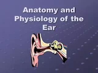
BASIC DIVISIONS OF EAR MIDDLE EAR INNER EAR EXTERNAL EAR
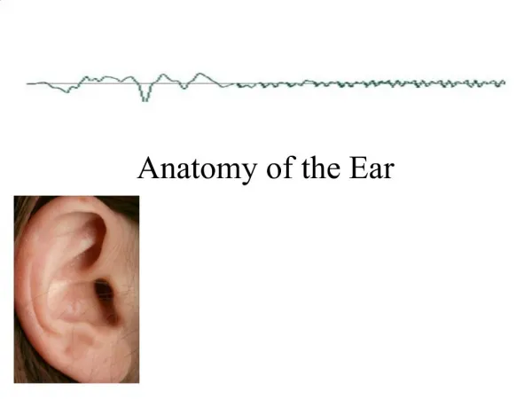
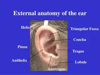
AURICLE Prominences & Depressions Ligaments
AURICULAR MUSCLES EXTRINSIC INTRINSIC • 3 in number • Auricularis Anterior • Auricularis Superior • Auricularis Posterior • 6 in number • Helicis major • Helicis minor • Tragicus • Antitragicus • Transversusauriculæ • Obliquusauriculæ
NERVE SUPPLY • Greater Auricular Nerve • Lesser Occipital Nerve • Auricular Nerve • Auriculotemporal Nerve • Facial Nerve
EXTERNAL AUDITORY CANAL • From Concha to T.M. • Length24mm • Cartilaginous-8mm • Bony-16mm • 2 Suture Lines • Tympanosquamous • Tympanomastoid • Medial End-Tympanic Sulcus
BLOOD SUPPLY OF E.A.C. • Auricular Br. Of Superficial Temporal Artery • Deep Auricular Br. Of Maxillary Artery {1st Part} • Auricular Br. Of Posterior Auricular Artery
MIDDLE EAR CLEFT • Tympanic Cavity • Eustachian Tube • Mastoid Air Cell System
TYMPANIC CAVITY • Compartments • Epitympanum • Mesotympanum • Hypotympanum • Boundaries • Lateral Wall • Roof • Floor • Anterior Wall • Medial Wall • Posterior Wall
COMPARTMENTS EPITYMPANUM MESOTYMPANUM HYPOTYMPANUM
THE LATERAL WALL • Wedge Shaped • 3 Divisions • Bony Lateral Wall Of Epitympanum • Tympanic Membrane • Bony Lateral Wall Of Hypotympanum • 3 Holes • Petrotympanic fissure • Anterior Canaliculus • Posterior Canaliculus
LATERAL WALL
TYMPANIC MEMBRANE • Forms lateral wall centrally • Oval Shaped • 55° Angle with floor • Longest Dia. • -10 mm(P.S.-A.I.) • Two Divisions • Pars Flaccida • Pars Tensa
3 LAYERS OF T.M. • Outer- Outer Epithelial Layer • Middle- Lamina Propria (Fibrous Layer) • Inner- Mucosal Layer • BLOOD SUPPLY OF T.M. • Epidermal vessels- Deep Auricular Br. Of Maxillary A. • Extensive anastomosis- In Lamina Propria • Mucosal vessels • Anterior Tympanic Br.- Maxillary A. • Stylomastoid Br.- Post. Auricular A.
COURSE & RELATIONS IN TYMPANIC CAVITY • FACIAL NERVE • LINGUAL NERVE
THE FLOOR • Jugular Wall • Thickness- Related to Jugular Fossa Height • Tympanic Br. Of CN IX HYPOTYMPANUM JUGULAR WALL JUGULAR BULB
THE ANTERIOR WALL • Carotid Wall • 3 Parts • Upper 1/3rd :- Canal Of Tensor Tympani, Anterior epitympanic sinus • Middle 1/3rd :- Tympanic orifice of Eustachian Tube • Lower 1/3rd :- Superior & Inferior Caroticotympanic N., Tympanic Br. Of ICA
Promontory • Basal Coil Of Cochlea • Tympanic plexus nerves • Oval Window • Behind & Above Promontory • Kidney Shaped • 3.25mm long • 1.75mm wide • Connects to Vestibule • Stapes footplate & Annular Lig.
Round Window:- • Below & behind Oval Window • Subiculum • Ponticulus • Triangular shaped • Sinus Tympani • Covering Membrane- • oval, 2.3 x 1.9mm
Facial Canal aka Fallopian Canal • Antero-posteriorly above Oval Window & Promontory • ProcessusCochleariformis—Anterior Landmark • Tensor Tympani tendon attachment
THE POSTERIOR WALL • Aditus Ad AntrumOpening to Mastoid Antrum • Fossa Incudis Contain Short Process Of Incus & It’s Suspensory Ligament • Pyramid Attaches Stapedius & It’s tendon
CONTENTS OF TYMPANIC CAVITY • Ossicles3 • Muscles2 • Chorda Tympani N. • Tympanic Plexus • Mucosal Folds • Blood Vessels
MUSCLES • Stapedius • Tensor Tympani
MUCOSAL FOLDS OF TYMPANIC CAVITY Mucus secreting respiratory mucosa with cilia 3 Mucociliary Pathways • Epitympanic • Promontorial • Hypotympanic
Mesotympanum & EpitympanumSeperated By TENSOR FOLDS INTEROSSEUS FOLD MEDIAL INCUDAL FOLD
Lateral View:- (13) Tensor Folds, (14) Anterior Malleolar Fold, (39) Tensor Tympani Tendon, (17) Interosseous Fold, (9) Medial Incudal Fold, (23) Superior Malleal Ligament, (11) Superior Incudal Fold, (24) Posterior Incudal Fold
Inferior View:Tensor Fold (13), The Interosseous Fold (17), The Medial Incudal Fold (9), Isthmus Tympani Anticus (20), Isthmus Tympani Oticus (21), (5)ObturatoriaStapedis, (8)PlicaStapedis; (11) Superior Incudal Fold, (14) Anterior Malleal Fold, (18) Superior Malleal Fold, (19)IncisuraTensoris, (22) Anterior Malleal Ligament, (23) Superior Malleal Ligament, (24) Posterior Incudal Ligament, (28) Pyramidal Eminence, (39) Tensor Tympani Tendon.
ISTHMUS TYMPANI • Narrow elongated space • Medially • Facial Nerve Canal • Lateral SCC • Divided by Long Process Of Incus • Isthmus Tympani Anticus • Isthmus Tympani Posticus
Prussack’s Space • Boundaries: • Medially: Neck Of Malleus • Laterally: Pars Flaccida • Inferiorly: Lateral Process Of Malleus • Superiorly: Lateral Malleolar Fold • Posteriorly: Posterior Malleolar Fold • Clinical Significance: • Pars flaccida acquired cholesteatoma formation
PATTERN OF SPREAD OF CHOLESTEATOMA • Posterior Epitympanum (Through Superior Incudal Space) • Posterior Mesotympanum (Through Posterior Pouch Of Von Tröltsch) • Anterior Epitympanum (Anterior to head of malleus to Supratubal recess)
EUSTACHIAN TUBE • Bony Part (Lateral 1/3rd) • 12mm , Rectangular opening • Cartilaginous (Medial 2/3rd) • 24mm • Torus Tubaris • Blood Supply • Ascending Pharyngeal A. • Middle Meningeal A.
Functions: • Pressure Equalization • MucociliaryClearance & Drainage • Protection of middle ear from nasopharyngeal environment & loud sounds
IMPROPER FUNCTIONING OF EUSTACHIAN TUBE Recurrent URTI, Enlarged Adenoids Larger Diameter & Shorter Length Malfunctioning of Tubal Muscles Reduction in Size Of Ostmann’s Fat Pad Atrophy Of Tensor VeliPalatini Collagen Diseases Loss Of Ciliated Epithelium Abonormally Thick & Tenacious Secretions