Download
1 / 23
230 likes | 401 Views
2. Anatomy Review. Bones of the regionfemur -- longest/strongest bone in the bodypatella -- sesamoid bonetibia -- largest bone in the lower leg; 2nd largest in bodyfibula carries approx 10% of body weightMuscles of the regionquadriceps (extension)hamstrings (flexion)abductors (abducts)adductors (adducts).

E N D
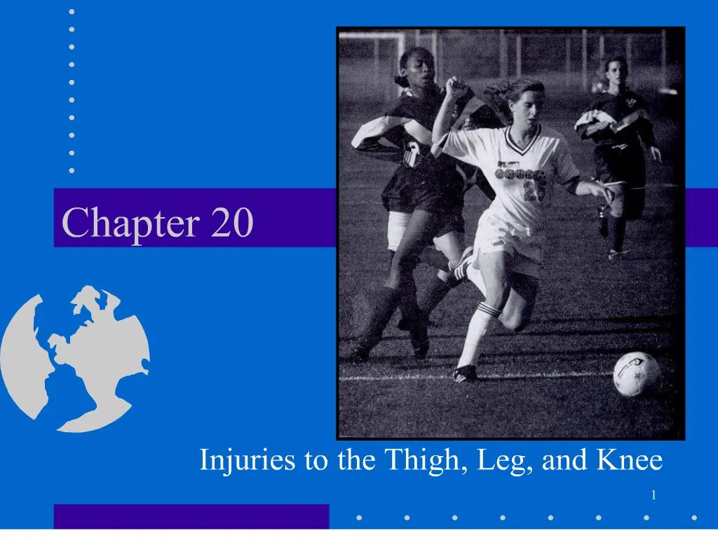
1. 1 Chapter 20 Injuries to the Thigh, Leg, and Knee
2. 2
3. 3 Anatomy Review Knee articulations
4. 4
5. 5 Anatomy Review
6. 6
7. 7 Common Sports Injuries - Bursitis Soft tissue injuries: bursa
small fluid-filled sac located at strategic points
numerous bursa around the knee region -- only a few are typically injured
inflammation can be caused by trauma, chronic irritation or infection
the prepatellar bursa is often irritated by trauma
8. 8 Common Sports Injuries - Bursitis Signs/symptoms:
swelling/tenderness
increased pain caused by manual pressure
history of trauma or chronic injury
First Aid:
apply ice and compression
reduction of activity
if chronic -- anti-inflammatory agents with a physician�s approval
9. 9 Common Sports Injuries � Patella Patellofemoral conditions
Some conditions of the patella may be directly or indirectly related to Q-angle.
As shown in the illustration, Q-angle is computed as the difference between a line drawn from the center of the patella compared with one drawn from the center of the patella through the center of the tibia.
Excessive Q-angle may be related to problems such as patellar chondromalcia or ACL tearing.
10. Proper Patellar Tracking 10
11. 11 Common Sports Injuries � Femur FX Signs & Symptoms
pain at the injury site
difficulty in walking
swelling and/or deformity
athlete may report having suffered a traumatic event
athlete may report a pop or snap at time of injury
First Aid:
splint the injured leg
apply sterile dressings to any related wounds
monitor vital signs and circulation of the lower leg
refer to a physician via ambulance
History
results from an extremely traumatic event
may also be in the form of a stress fracture, especially the femoral neck region
In the adult, fractures of the femoral neck may result in avascular necrosis of the femoral head.
This injury results from disrupted blood supply to the articular cartilage on the femoral head.
12. 12 Common Injuries - Muscle Strains History
Sudden acceleration or deceleration (acute)
Fatigue induced (chronic)
May report feeling/hearing pop or snap
Sign/symptoms
A sharp pain in the affected muscle
Swelling
weakness & inability to contract the muscle forcefully
discoloration (2nd degree)
visible deformity (3rd degree) First aid
I.C.E.
rest
anti-inflammatory
refer to a physician (3rd degree)
13. 13 Common Sports InjuriesPatellar Dislocation/Subluxation History
Onset: Acute or chronic
Location of Pain: Medial jt capsule as well as pain under the patella.
Mechanism: forceful extension with femoral ext rotation while knee is in flex
Observation:
Unreduced patellar show an obvious deformity
Swelling within 24 hrs after onset of injury
Palpation
Pain over the lateral & medial side of the patella
Origin of the VMO
Along the Patella Tendon Functional
AROM: Pain during the 1st 300 of flexion or terminal extension
RROM: decrease isometric str. During extension when knee is flexed 00 & 300.
Special Test
(+) Apprehension Test [subluxation]
14. 14 Common Sports InjuriesPatellar Dislocation/subluxation Treatment:
slowly extend leg (reduce)
I.C.E
immobilize the injury with knee immobilizer for 4 weeks
treat any open wounds with sterile dressings
monitor vital signs and circulation of the lower leg
Refer to MD
15. 15 Patellar Tendonitis �Jumpers Knee� History
Onset: Chronic
Location of Pain: Inferior pole and/or insertion site at the tibial tuberosity or tendon itself.
Mechanism: Repetative movement involving resisted knee extension
Observation
Swelling around the patellar tendon & inferior pole of the patella or tibial tuberosity (Osgood-Schlatter)
Palpation
Tenderness on the patellar tendon & inferior patella pole or tibial tuberosity Functional
AROM: Pain during knee extension
PROM: Pain during end of knee flexion
RROM: Pain throughout knee flx ROM
First Aid:
Ice
NSAIDs
Rest
Patella strap
16. 16 Common Sports InjuriesMyositis Ossificans Soft tissue injuries
contusions -- can occur anywhere but are common in the anterior thigh --
sports such as football and ice hockey require padding of the region for prevention
if force of injury is sufficient a deep bruise may result -- including damage to the underlying periosteum
if not cared for properly, myositis ossificans may develop Signs/symptoms:
history of forceful impact
muscle spasm and swelling
loss of function & pain
difficulties in walking
First Aid:
apply ice and compression
if severe, put on crutches
rest
avoid further irritation until the acute phase has passed
protective doughnut padding with a thermoplastic dome required when athlete returns to play
17. 17 Common Sports InjuriesMeniscus tears History
Onset: Acute
Location of pain:Along the medial or lateral jt line
Mechanism: Tibal rotation w/ flexion & valgus or varus force.
Observation
Jt effusion may develop over 24 to 48 hrs. In the JT line or popliteal fossa
Palpation
Pain along the JT line and b/w the joint line when knee is flexed to 90�
Functional
AROM: ROM is decreased
PROM: Pain at extreme of flexion/extension
RROM: Pain or locking is revealed
Special Test
(+) McMurray�s test
(+) Apley�s Compression
Note: Test ACL & MCL
First Aid:
ice and compression
put athlete on crutches
refer to a physician
18. 18 Common Sports Injuries-MCL/LCL Sprain History
Onset: Acute
Location of pain: Medial side of the knee (MCL), Lateral side of the knee & fibula head (LCL)
Mechanism: Valgus force or excess external rotation (MCL), Varus force or excess interal rotation (LCL)
Observation
Swelling medial side (MCL), lateral side (LCL)
19. 19 Common Sports Injuries-MCL/LCL Sprains Palpation
Tenderness along the length of the MCL or LCL
Functional
AROM: pain on terminal ranges of flexion & extension (MCL/LCL)
PROM: pain on terminal ranges of flexion & extension (MCL/LCL)
RROM: decreased strength & pain during flexion/extension Special test
Valgus stress test (MCL)
Varus stress test (LCL)
First Aid:
ice and compression
Immobilize PRN
put athlete on crutches PRN
Note: 3o refer to doctor
20. 20 Common Sports Injuries-ACL/PCL History
Onset: Acute
Location of pain: within the knee joint radiating anteriorly (ACL) or posteriorly (PCL)
Mechanism: (ACL) tibia moves anterior on the femur or femur moves posterior to tibia. Hyperextension of the knee. Rotation also tears ACL . Most are noncontact. (PCL) tibia moves posterior to femur when @ 90� angle. Requires some sort of force . Aka Dashboard injury
Observation
Rapid swelling
Tibia sagging (PCL) Palpation
(ACL) cannot palpate
(PCL) tenderness in the poplital fossa
Functional
(ACL) AROM: swelling may prohibit full ROM
PROM: pain throughout the ROM
RROM: if pain on AROM & PROM, do not perform RROM
(PCL) AROM: pain when fully flexed
PROM: pain when nears full flexion
RROM: pain near terminal flexion
21. 21 Common Sports Injuries-ACL/PCL Special Test
(ACL) Anterior Drawer test
(ACL) Lachman�s test
(PCL) Godfrey�s sign
(PCL) Posterior Drawer test
22. 22 ACL � Surgical Options ACL Repair
Suturing of torn ACL to itself
Highly unsuccessful; outdated
ACL Reconstruction
Patellar tendon or Hamstring (Semitendinosus)
Patellar used to be favored, but slightly longer rehab intially & pain b/c of tendon
Semitendinosus causes less pain but hamstring can bother later
Cadaver � ACL or Achilles
23. 23 ACL Rehab Rehab lasts anywhere from 4-6 mons
ROM is most important along with protection of graft
No open chain resisted knee ext til at least 4 mons
Mechanical issues must be addressed; particularly take-off and landing
Hamstrings must also be strengthened
24. ACL Prevention Women are 5x more likely to tear their ACL
Hormones?
Higher Q-angle
Lack of hamstring recruitment
Improper take-off/landing technique
Prevention Programs Developed
SportsMetrics � �knees under the hips and over the ankles�
6 week program
Incorporates flexibility, strength, and plyometric training
Reinforces proper take-off and landing techniques
Not only reduces likelihood of ACL tear but also improves athletic performance. 24