Download
1 / 10
100 likes | 107 Views
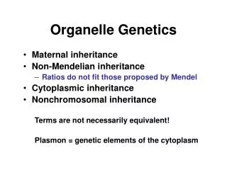
Subcellular Organelle structure service for students,scholars,researchers...

E N D
Cryo-Electron Microscopy (Cryo-EM) can present samples in a native state with imaging the sample’s intrinsic electron density, providing direct visualization of biological macromolecules, such as protein, nucleic acid, large complexes and so on. Conversely, X-ray crystallography must require crystallization of the specimen within non-physiological environments. The resolution of Cryo-EM maps has been improved up to near-atomic resolution level. Thus Cryo-EM is gaining popularity in structural biology.
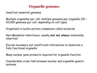
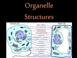
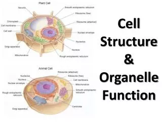
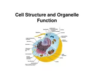
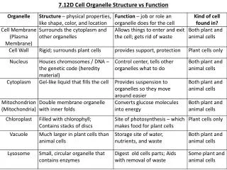
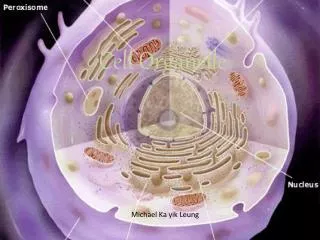
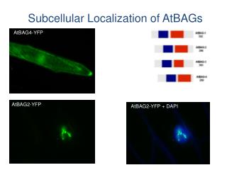
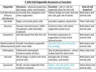
Cryo-Electron Tomography (Cryo-ET), one of the techniques of Cryo-EM, is used to produce three-dimensional (3D) pictures from a series of tilted Cryo-EM images. Cryo-ET has gained more and more attention in the field of Cryo-EM in the recent years, which can be applied to study the internal organisms, typically for many structures inside cells, for example, subcellular organelles. Organelle is a specialized subunit within a cell, which contains cytoskeleton, mitochondria, endoplasmic reticulum, flagellum and so on. Cryo-EM allows to study the high-resolution structures of macromolecules, and Cryo-ET provides the information about cellular processes. With the combination of 3D imaging and specimen preparation that preserves the integrity of the cell structure, Cryo-ET can observe the internal structure of cell with high resolution.
At Creative Biostructure, we have the most advanced Electron Microscopes equipments and software. In addition, Creative Biostructure has a professional team for experimental design of subcellular organelles separation and Cryo-ET data analysis.
The advantages of our service include: The most advanced direct-electron detectors and image processing software; Allow near-atomic resolution structures to be calculated without crystallization and only need a small amount of purified samples; Provide the unique possibility to image organelle in their native environment or in situ; Visualize the interaction of proteins and cells, capture transient and dynamic processes; Bring a hope for further understanding of life mechanisms and mysteries.
Creative Biostructure provides professional services of Cryo-EM. It is possible to observe the structure of cells in the native state and provide the detailed information of life.
References • Muyuan Chen, Wei Dai, et al. Convolutional neural networks for automated annotation of cellular cryo-electron tomograms. Nat Methods, 2017; 14:983-985. • Takashi Ishikawa. 3D structure of eukaryotic flagella/cilia by cryo-electron tomography. Biophysics (Nagoya-shi), 2013; 9:141-148. • Tanmay A.M. Bharat, Sjors H.W. Scheres. Resolving macromolecular structures from electron cryotomography data using sub-tomogram averaging in RELION. Nat Protoc, 2016; 11: 2054-2065.
Excerpt from: https://www.creative-biostructure.com/Cryo-EM-for-Subcellular-Organelles-598.htm