Download
1 / 1
10 likes | 124 Views
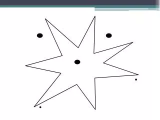
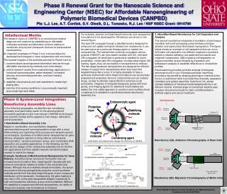
Other Nanofactories. M. M. Fastest migraters. Least adhesive. Expansion chamber A. Expansion chamber A. Primary cells. ‘Cells’. Expansion chamber B. Expansion chamber B. Slowest migraters. Most adhesive. Gradually increasing magnetic field. Expansion chamber C.

E N D
Other Nanofactories M M Fastest migraters Least adhesive Expansion chamber A Expansion chamber A Primary cells ‘Cells’ Expansion chamber B Expansion chamber B Slowest migraters Most adhesive Gradually increasing magnetic field Expansion chamber C Expansion chamber C Mobile trap array T-cells, glioma cells, primary cells Three new polymer and lipid-based formulations. Schematic of a nanovector design for delivery of siRNA. Phase II Renewal Grant for the Nanoscale Science and Engineering Center (NSEC) for Affordable Nanoengineering of Polymeric Biomedical Devices (CANPBD)PIs: L.J. Lee, A.T. Conlisk, S.V. Olesik, D.L. Tomasko, R.J. Lee / NSF NSEC Grant: 0914790 Intellectual Merits For example, polymer and lipid-based molecular and nanoparticle formulations to be developed for ON delivery are shown in the second figure. The lipid-ON conjugate through a disulfide bond is designed for enhanced cell uptake and quick release from endosomes. It can be used alone as a molecular therapy agent or loaded into nanoparticles. The hydrophobic molecule conjugated cationic polymer (HMCCP) can form stable micelles with ON. Like lipid-ON conjugates, its small size is particularly valuable for tumor penetration. Unlike lipid-ON conjugates, micelles allow higher ON loading. Again, they can be loaded in nanoparticles for delivery. The two-stage liposomal nanoparticles are designed for efficient release of cargos from nanoparticles when they reach the targeted tissue like a tumor. It is highly desirable to have a synthesis method with which these formulations can be precisely prepared and evaluated. As more components such as multiple drugs, nuclear localization signal (NLS) to enhance nuclei permeation, integrase to integrate therapeutic genes with host genes, and imaging agents for additional functionalities are added, the only viable approach to construct such multifunctional complexes is to establish a nanofluidics-based nanofactory assembly line. 2. Nanofiber-Based Nanofactory for Cell Separation and Analysis The second nanofactory integrates a foundation of electrospun nanofiber with post-processing using femtosecond laser ablation and supercritical fluid-based impregnation. The figure below shows an example of cell separations that can occur: 2(A) stem cell separation into different expansion chambers based on relative adhesion into patterned microwells and magnetic manipulation; 2(B) migratory cell separation on aligned nanofiber arrays followed by expansion and subsequent analysis to establish differences in intracellular profiles. Electrospinning provides synthetic analogs of biological structures found in vivo. Femtosecond laser machining provides a key benefit by shaping electrospun matrices at the micron or submicron level to provide flow channels, separation chambers and guided assembly. Subcritical CO2 allows ‘printed’ additions of chemical functionality/bioactivity in an efficient manner. A broad range of envisioned medical uses includes microenvironments for stem cell differentiation, artificial organs and cancer treatment. • The research vision of CANPBD is to revolutionize medical diagnosis and medicine by establishing an affordable multiscale synthesis and fabrication protocol leading to nanofluidic and polymer therapeutic devices for personalized nanomedicine. • An important emphasis of Phase II is to commercialize the developed technologies in close collaboration with end users. • The broader impacts of the activities planned for Phase II are to: • commercialize nanoengineered biomedical devices through affordable manufacturing methods and novel design; • extend research results from medical/biology applications to functional nanocomposites, water treatment, homeland security, environmental protection, and food industry toxicology; • establish new products and new industries to create high-paying jobs; • train the 21st century workforce in economically important and critical high-tech fields. • Phase II System-Level Integration: • Nanofactory Assembly Lines • In the following paragraphs, we describe two nanofactory assembly lines particularly useful for the aforementioned biomedical applications and how relevant CANPBD technologies and scientific studies will be applied to their design, fabrication, and bioevaluation. • Nanofluidics-Based Assembly Line • Based on nanofluidics, one nanofactory integrates nanomanufacturing and nanomanipulation arrays with a novel DNA combing and imprinting (DCI) process and nanowire electric circuit designs. Synthesis of multifunctional nanoparticles for gene delivery, drug/gene injection to living cells by nanochannel electroporation, nanowire biosensor arrays, and nanofluidics DNA separation are possible applications. In the following, we first describe the design of this nanofactory assembly line for the first two applications and then explain relevant nanotechnologies needed to realize the design. • Example: Synthesis of Multifunctional Nanoparticles for Gene Delivery. A multifunctional nanovector formulation that can increase blood circulation time, target specific tissues/cells and enhance intracellular release is shown in the schematic that follows. Although this is a relatively simple formulation, the actual structures of forming nanoparticles using the current synthesis methods are far from this ideal shape because of poor component distribution at the nanoscale. Consequently, the gene loading is very low (<10% of the total nanoparticle weight), cytotoxicity is high and transfection efficiency is poor and inconsistent. Without the capability to prepare well-defined nanoparticles, our ability to design and evaluate new formulations is hindered. Nanofactory 2(A): Adhesion Chromatography of Stem Cells Nanofactory 2(B): Migratory Chromatography of Motile Cells This material is based on work funded by the National Science Foundation (Grant # 0914790)