Download
1 / 31
410 likes | 1.49k Views
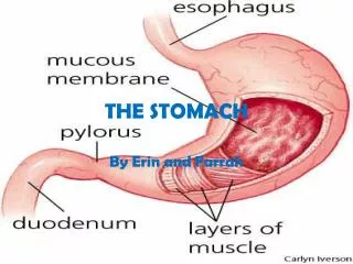
Tumors of the stomach and duodenum. Benign Polyps Hyperplastic Fundic gland Neoplastic Multiple Tumors Leiomyomas Lipomas Heterotopic pancreas. Malignant Tumors Carcinoma Lymphoma Sarcoma Carcinoid Others Menetriers Disease Bezoar Volvulus. INTRODUCTION - STOMACH.

E N D
Benign Polyps Hyperplastic Fundic gland Neoplastic Multiple Tumors Leiomyomas Lipomas Heterotopic pancreas Malignant Tumors Carcinoma Lymphoma Sarcoma Carcinoid Others Menetriers Disease Bezoar Volvulus INTRODUCTION - STOMACH
GASTRIC POLYPS • Hyperplastic polyps • Most common type of polyp (65 – 90%) • Inflammatory or regenerative polyps • In reaction to chronic inflammation or regenerative hyperplasia • Often found in HP infections • Sessile and seldom pedunculated • Mostly in the antrum • Multiple in 50% of cases • Varying in size but seldom < 2cm • Rate of malignant transformation 1 – 3% • Usually larger than 2 cm
GASTRIC POLYPS • Fundic Gland • Small elisions in the fundus • Hyperplasia of the normal fundic glands • Often associated with FAP • Therefore important as a marker for disease elsewhere in the GIT tract
GASTRIC POLYPS • Neoplastic polyps • Types • Tubular • Villous (often larger - > 2cm - and malignant) • Macroscopically • More often in antrum • Pedunculated with malignant potential • Solitary, large and ulcerated • Treatment • Endoscopic removal if no malignancy identified with surveillance • Excision with malignant focus or where endoscopic removal failed
GASTRIC POLYPS • Multiple gastric polyps • Rare condition • Adenomatous and hyperplastic polyps • 20% incidence f adenocarcinoma • Treatment • If confined to corpus and antrum – distal gastrectomy • Otherwise total gastrectomy • Sometimes associated with Polyposis syndromes • FAP • Gardner • Peutz-Jeghers • Cowden • Cronkhite Canada
GASTRIC LEIOMYOMA • Incidence of 16% at autopsy • Pathology • Arise from smooth muscle of the GIT tract • Difficult to distinguish from GIST • 75% benign • Differentiation only on mitotic index • Large protruding elisions with central ulcer • Usually presents with bleeding if at all • Treatment is local excision with 2 – 3cm margin
GASTRIC LIPOMA • Rare subcutaneous lesions • Asymptomatic • On routine endoscopy • Require no treatment • Pillow sign
HETEROTOPIC PANCREAS • Ectopic pancreas • Most common found in stomach • Within 6 cm from the pylorus • Also in Meckl’s diverticulum • Rarely larger than 4 cm • Sessile and rubbery • Submucosal in location • Histological identical to normal pancreas
ADENOCARCINOMA OF THE STOMACH • Declining incidence in western world • HP associated due to chronic atrophic gastritis • Also related to • Low dietary intake vegetables and fruit • High dietary intake of starches • More common in males ( 3 : 1 ) • Histology • Invariably adeno-carcinoma • Squamous cell carcinoma from oesophagus • Involves fundus and cardia
ADENOCARCINOMA OF THE STOMACH • Histological typing • Ulcerated carcinoma (25%) • Deep penetrated ulcer with shallow edges • Usually through all layers of the stomach • Polipoid carcinoma (25%) • Intraluminal tumors, large in size • Late metastasis • Superficial spreading carcinomas (15%) • Confinement to mucosa and sub-mucosa • Metastasis 30% at time of diagnosis • Better prognosis stage for stage
ADENOCARCINOMA OF THE STOMACH • Histological typing • Linitis plastica (10%) • Varity of SS but involves all layers of the stomach • Early spread with poor prognosis • Advanced carcinoma (35%) • Partly within and outside the stomach • Represents advanced stage of most of the fore mentioned carcinomas
ADENOCARCINOMA OF THE STOMACH • Symptoms and signs • Vague discomfort difficult to distinguish from dyspepsia • Anorexia • Meat aversion • Pronounced weight loss • At late stage • Epigastric mass • Haematemesis usually coffee ground seldom severe • Metastasis • Vircho node in neck • Blumer shelf in rectum
ADENOCARCINOMA OF THE STOMACH • Surgical resection only cure • Late presentation makes sugary often futile • Palliation controversial for • Haemorrhage • Gastric outlet • Simple gastrectomy as effective as abdominal block • Splenectomy often added due to direct involvement • Only for the very distal partial gestrectomy • Rest total gastrectomy • Prognosis overall 12% 5 year survival • 90% for stage I disease
GASTRIC LYMPHOMA • 5% of all primary gastric neoplasm's • 2 different types of lymphoma • Part of systemic lymphoma with gastric involvement (32%) • Part of primary involvement of the GIT (MALT Tumors) • 10 – 20% of all lymphomas occur in the abdomen • 50% of those are gastric in nature • Risk factors • HP due to chronic stimulation of the MALT • In early stages of disease Rx of HP leads to regression of the disease
GASTRIC LYMPHOMA Primary MALT • Early stages also referred to as pseudo-lymphoma • Indolent for long periods • Low incidence of • Spread to lymph nodes • Involvement of bone marrow • Therefore much better prognosis • Mostly involves the antrum • 5 different types according to appearance • Infiltrative - Ulcerative • Nodular - Polypoid • Combination
GASTRIC LYMPHOMA Primary MALT • At time of presentation • Larger than 10 cm (50%) • More than 1 focus (25%) • Ulcerated (30 – 50%) • Pattern of metastasis similar to gastric carcinoma • Signs and symptoms • Occur late and are vague • Relieved by anti-secretory drugs • Diagnosis based on histology
GASTRIC LYMPHOMA Primary MALT • Treatment controversial • Surgical treatment for patients without systemic involvement • Mandatory for high grade lesions • Possible not needed for low grade lesions • Total gastrectomy and en-block for direct involvement • Sparing duodenum and oesophagus • Palliative resection with intra-abdominal spread • Good for bleeding, obstruction and perforations • Radiation and chemotherapy combination for most
GASTRIC SARCOMA • 1 – 3 % of gastric malignancies • Include a wide variety of tumors • Leiomyosarcoma • Leiomyoblastoma • GIST
MENETRIERS DISEASE • Giant gastric folds (hypertrophic gastropathy) • Differentiate from • Infiltrating neoplasm (Ca / lymphoma) • CMV infection • Manifestation • Hypo-proteinaemia due to loss from ruggae • Chronic blood loss • Treatment • Medical (PPI, atropine, H2 blockers) • Surgical for refractory cases or where Ca cant be excluded
GASTRIC BEZOAR • Concretions in the stomach • Tricho-bezoar (hair) • Young girls who pick and swallow their hair • Phyto-bezoar (vegetable fibre) • Can cause erosions and bleeding • Seldom perforate but if mortality 20% • Post-gastrectomy predisposes • Both mechanical and chemical • Endoscopic breakage
GASTRIC VOLUVLUS • 2 Types • Organo-axial • Through the organs longitudinal axis • More common and associated with hiatus hernia • Eventration of the diaphragm • Mesenterio-axial • Line through mid lesser to mid greater curvature • Clinical triade (Brochardt’s) • Vomiting followed by retching and inability to vomit • Epigastric distension • Inability to pass NGT
GASTRIC VOLVULUS • Treatment • Emergency surgery as any volvulus
GASTRIC DIVERTICULAE • True diverticulae uncommon • Involve all layers of the wall • Pre-pyloric in location • Pulsion with only mucosa and sub-mucosa • Within a few cm of GEJ • Asymptomatic found on routine investigations • Confused with peptic ulceration
Benign Brunners gland adenoma Leiomyoma Carcinoid Heterotopic gastric mucosa Villous adenoma Malignant Peri-ampullar adeno CA Duodenum Cholangio Pancreatic head Leiomyosarcomas Lymphomas Others Duodenal dIverticula INTODUCTION - DUODENUM
DUODENUM Benign tumors • Brunners gland adenomas • Small submucosal • Sessile and pedunculated variants • Posterior wall junction D1 and D2 • Symptoms due to bleeding or onstruction • Leiomyoma • Asymptomatic • Carcinoid • Mostly active (gastrin, SS and serotonin) • Simple excision
DUODENUM Benign tumors • Hetrotopic gastric mucosa • Multiple small mucosal lesions • No clinical significance • Villous adenoma • Intestinal bleeding • Obstruction of ampulla with jaundice • Risk of malignancy high (50%) • Endoscopic snaring or local excision
DUODENUM Malignant tumors • Located in the descending part of the duodenum • Symptoms • Pain, obstruction bleeding and jaundice • Earlier than pancreas head • Treatment • Pancreatico-duodenectomy for localized lesions • Much better prognosis than pancreas Ca (30% 5-year as opposed to 0%) • Palliative bypass procedures if not resectable • Radiotherapy for advanced disease ?
DUODENAL DIVERTICULAE • Incidence • 20% at autopsy • 5 – 10% at upper GIT investigations • Pulsion diverticulae • 90% on the medial border of the duodenum • Solitary and within 2.5 cm of the ampulla • Associated gallstones and gallbladder disease • Pseudo-diverticluae • First part of the duodenum • Scarring of PUD
DUODENAL DIVERTICULAE • Presentation • Chronic post-prandial pain and dyspepsia • With complicated disease • Bleeding and perforation • Panceatitis • Jaundice • Surgery for complicated disease • Dissection, removal and closure (even with perforation) • With billiary involvement : cholidocho-duodenostomy