Download

1 / 7
70 likes | 91 Views
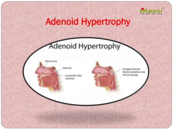
Adenoid is a subepithelial collection of lymphoid tissue situated at the junction of the posterior wall of the nasopharynx. It is also known as nasopharyngeal tonsil, having vertical ridges of lymphoid tissue separated by deep clefts.

E N D
WHAT IS AN ADENOID? Adenoid is a subepithelial collection of lymphoid tissue situated at the junction of the posterior wall of the nasopharynx. It is also known as nasopharyngeal tonsil, having vertical ridges of lymphoid tissue separated by deep clefts. It is covered by three epithelium i.e., transitional, pseudostratified ciliated columnar, and stratified squamous. Having no crypts and capsules. It is included in the waldeyer’s ring of lymphoid tissue. It is present at birth but shows physiological enlargement up to 6 yrs and then starts atrophy at puberty. It disappears by age of 20. It is seen in MRIs of all infants by the age of 5 months. The persistence of adenoids can be seen beyond the age of 15 years in case of allergy or infection. One in number Lymphatics from this tissue drain into upper jugular nodes directly or indirectly through retropharyngeal and Para pharyngeal lymph nodes. Arterial supply from ascending palatine branch of the al artery, ascending pharyngeal branch of the external carotid artery, ascending branch of the third part of the maxillary artery, and inferior thyroid artery’s ascending cervical branch. Sometimes inflammation of the adenoid lead to pain in the ear due to the same nerve supply as cranial nerves IX and X (referred pain) or can be due to eustachian tube obstruction.
ADENOID HYPERTROPHY It is an obstructive condition due to the enlargement of the adenoid which can be physiological enlargement in childhood or due to some infections or allergic conditions. WHAT IS THE ETIOPATHOGENESIS OF HYPERTROPHY? Recurrent sinusitis, rhinitis, or chronic infection of tonsils lead to chronic adenoid infection and hyperplasia of lymphoid tissue which results in adenoid hypertrophy. Allergy to the upper respiratory tract may lead to the enlargement of adenoids. Viral infections like EBV or bacterial infection of group A streptococci can lead to its enlargement. Exposure to smoking or pollution. Tumours of sinuses, lymphoma, and AIDS Due to diseases like GERD, there is a reflux of acid that will irritate the adenoid tissue leading to inflammation and resulting in hypertrophy.
HOW PATIENTS WILL PRESENT TO YOU IN OPD? Signs and Symptoms These depend not only on the size of the adenoid mass but also on the space that is available for enlargement in the nasopharynx. Excessive hypertrophy of the adenoid can lead to obstruction of the whole nasopharynx. In cases of enlargement, the patient may be asymptomatic but in severe enlarged and infected adenoids, the patient presents nasal, aural, and general symptoms. Nasal Symptoms The commonest symptom is nasal obstruction due to recurrent sinusitis and this leads to mouth breathing. Nasal obstruction leads to interference in the suckling or feeding of a child. As respiration and feeding cannot occur together, the child fails to thrive as all his efforts go into breathing which will lead to suboptimal growth of the child. There will be nasal discharge due to choanal obstruction as the normal secretion cannot drain back into the nasopharynx and is associated with chronic maxillary sinusitis and the child has a wet bubbly nose. When adenoids are acutely inflamed, epistaxis can occur when the patient blows his nose. Voice change can be seen i.e. toneless and losing nasal quality due to nasal obstruction and there will be rhinolaliaclausa i.e. hyponasal or denasal speech due to lack of appropriate nasal airflow during speech. Aural Symptoms Tubal Obstruction: Adenoid mass blocks the eustachian tube leading to retraction of the tympanic membrane and conductive hearing loss. Recurrent attacks of acute otitis media may occur due to the spread of infection through the eustachian tube. CSOM may fail to resolve in presence of infected adenoids. There will be fluctuating hearing loss due to differences in the size of the mass of the adenoid. Impedance audiometry test helps to identify fluid in the ear due to the presence of glue ear that is otitis media with effusion. General Symptoms Adenoid Facies: Chronic nasal obstruction and mouth breathing lead to characteristic facial appearance. Dull expression because he is unable to hear and elongated face, open mouth, prominent and crowded upper teeth, hitched up upper lip, pinched up nose because the child is a mouth breather so there will be no use of nose for a long time for breathing that will lead to disuse atrophy of alar prominence, nasal crease and labial folds will be absent, high arched palate because the child is breathing through the mouth and as the mouth is continuously open, more air goes through the palate. Pulmonary hypertension: As the child snores a lot during the night and there is sleep apnoea (episodes of not breathing during sleep), the child gets up again and again during the night and is unable to have a good night’s sleep. This results in sleepiness and lethargy throughout the day. In sleep apnoea, so many episodes of deoxygenation happen that the pulmonary artery undergoes vasoconstriction which leads to pulmonary hypertension and results in right-sided heart failure. The final consequence is corpulmonale. Aprosexia i.e. lack of concentration in studies.
How can the diagnosis be done? Posterior Nasal Rhinoscopy: Examination of postnasal space in young children using a mirror from which adenoid mass can be visualized. Nasal endoscopy: Done with the help of a rigid or flexible nasopharyngoscope which helps in assessing the size of adenoid mass from which grading can be done. It is done on cooperative children. It provides more details on the nasopharynx. Soft tissue lateral radiograph of the nasopharynx: Done if the child is uncooperative as it will reveal the size of the adenoid and also the extent to which nasopharyngeal air space is compromised. There will be very little breathing space in the nasopharynx in severely enlarged mass.
FAQs (Frequently asked questions) Q1. How an adenoid hypertrophy patient will present to you? Ans. The child’s mouth would have been open for the past few weeks and he might not be responding. It would be seen only when the season changes or if the child is allergic to any substance. Q2. Why an adenoidectomy is contraindicated in velopharyngeal insufficiency? Ans. In velopharyngeal condition, the child is supposed to have regurgitation of food from the nose and hyponasal speech post-surgery so it is not advised to perform adenoidectomy in such patients. Q3. How adenoid hypertrophy can be differentiated from an antrochoanal polyp? Ans. In polyp, the crescent or Dodd sign is present whereas it is absent in adenoid hypertrophy.
Let’s Connect for More Support: care@diginerve.com Enquiries: +91 8800 418 418 https://www.diginerve.com/ https://www.facebook.com/diginerve.jaypee/ https://www.instagram.com/diginerve.jaypee/ https://twitter.com/diginerve