Download
1 / 43
430 likes | 1.04k Views
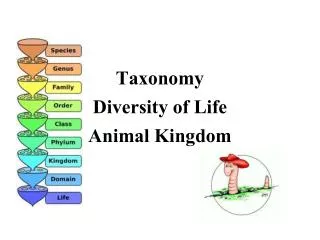
An introduction to the diversity of animal life. One cell or many?. We start dividing up animals here. Some animals have just one cell – many others have large numbers of differentiated cells. 1 cell - Protozoa. Many cells – parazoa and metazoa. The Protozoa – the single celled animals.

E N D
One cell or many? We start dividing up animals here. Some animals have just one cell – many others have large numbers of differentiated cells. 1 cell - Protozoa Many cells – parazoa and metazoa
The Protozoa – the single celled animals In fact many of these are photosynthetic and are claimed as plants by botanists, while some are both photosynthetic and carnivorous! The animal -plant - fungus split does not make sense at this level. Old system: exclude green species, lump the rest in Phylum protozoa, which has 4 classes: ciliates (Paramecium caudatum) – many small cilia flagellates (Euglena, Trypanosoma) – one big cilium (flagellum) Rhizopoda (Amoeba proteus) – no cilia + a less well known class of parasitic species: Sporozoa (Plasmodium vivax)
Ciliates are covered in hundreds of tiny motile hairs = cilia (sing. cilium). Are common in freshwater, also benign gut inhabitants. flagellates move by a small number of long motile hairs = flagellae (sing. flagellum). Free living, also rumen flora and some gut parasites. Rhizopoda free living in sediments etc, moving by slow protrusion of pseudopodia. A few are nasty parasites (Entamoeba dysenterica, Naegleria spp.) Sporozoa (Plasmodium vivax causes malaria, the biggest killer in human history)
New version – kingdom Protozoa Instead of the drastic shoe-horning described above, the current version is to regard all single-celled organisms as belonging to the kingdom Protozoa with many phyla (27 at last count!) This is probably more realistic, but much harder to remember.
Sponges – Phylum parazoa These are essentially colonial protozoa, whose colonies are reinforced with solid spicules of various shapes and composition. Silica SiO2 and Calcite CaCO3 are the commonest. They are exclusively aquatic, mainly marine, and live by filter feeding. The feeding cells are called choanocytes, which incorporate a central flagellum pumping water through the sponge, and the water passes through a collar of cilia-like filtering projections. The other main cell type is ameoba-like, making the supporting tissues and moving nutrients around. Typically sponges suck water in from around their bodies and exhale it from a common central siphon. Due to their diffuse form, and often variable colour, identifying them is often difficult / impossible in the field and relies on microscopic examination of spicules.
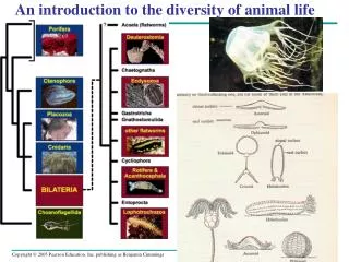
Metazoa: These are animals with fully differentiated tissues, including muscles and nerves. Many cells 1 cell - Protozoa No clear tissues: parazoa Tissues: metazoa The next level up in organisation takes us to the group of animals that used to be classed as phylum coelenterata (jellyfish, anemones and sea gooseberries). These are now split into 2 phyla, based on deep differences in design of their their stinging cells: Cnidaria – jellyfish and anemones Ctenophora – sea gooseberries.
Bilateria: this comprises c. 25 phyla all with bilateral symmetry (at least as larvae) and 3 layers of cells in the embryo. Many cells 1 cell - Protozoa No clear tissues: parazoa Tissues: metazoa Radial symmetry 2 cell layers in embrya Phyla cnidaria and ctenophora Bilateral symmetry 3 cell layers in embryo Remaining animal Phyla
The big 5 coelomate phyla There are about 10 phyla in which the basic body design involves a body cavity lined with cells (called a coelom), but of these I will only cover 4 today – these are the important common ones. One grouping is probably 3 distantly related phyla. Phylum annelida – the segmented worms Phylum mollusca: snails and allies Phylum echinodermata – starfish and allies Phylum (superphylum?) arthropoda – insects, spiders and crustaceans. Phylum chordata – everything with a backbone (including us)
Protostome and Deuterostome Development • Based on certain features seen in early development • Many animals can be categorized as having one of two developmental modes: protostome development or deuterostome development
Deuterostome development (examples: echinoderms, chordates) Protostome development (examples: molluscs, annelids, arthropods) (a) Cleavage. In general, protostomedevelopment begins with spiral, determinate cleavage.Deuterostome development is characterized by radial, indeterminate cleavage. Eight-cell stage Eight-cell stage Spiral and determinate Radial and indeterminate Cleavage • In protostome development • Cleavage is spiral and determinate • In deuterostome development • Cleavage is radial and indeterminate Figure 32.9a
(b) Coelom formation. Coelom formation begins in the gastrula stage. In protostome development, the coelom forms from splits in the mesoderm (schizocoelous development). In deuterostome development, the coelom forms from mesodermal outpocketings of the archenteron (enterocoelous development). Coelom Archenteron Coelom Mesoderm Blastopore Mesoderm Blastopore Enterocoelous: folds of archenteron form coelom Schizocoelous: solid masses of mesoderm split and form coelom Figure 32.9b Coelom Formation • In protostome development • The splitting of the initially solid masses of mesoderm to form the coelomic cavity is called schizocoelous development • In deuterostome development • Formation of the body cavity is described as enterocoelous development
Mouth Anus Digestive tube Anus Mouth Mouth develops from blastopore Anus develops from blastopore Figure 32.9c Fate of the Blastopore • In protostome development • The blastopore becomes the mouth • In deuterostome development • The blastopore becomes the anus
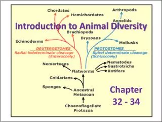
Leading hypotheses agree on major features of the animal phylogenetic tree • Zoologists currently recognize about 35 animal phyla • The current debate in animal systematics • Has led to the development of two phylogenetic hypotheses, but others exist as well
Rotifera Cnidaria Porifera Annelida Mollusca Chordata Phoronida Nemertea Ctenophora Nematoda Arthropoda Ectoprocta Brachiopoda Echinodermata Platyhelminthes “Radiata” Deuterostomia Protostomia Bilateria Eumetazoa Metazoa Ancestral colonial flagellate • One hypothesis of animal phylogeny based mainly on morphological and developmental comparisons Figure 32.10
Cnidaria Chordata Mollusca Annelida Rotifera Silicarea Phoronida Nemertea Calcarea Arthropoda Ctenophora Ectoprocta Brachiopoda Nematoda Echinodermata Platyhelminthes “Radiata” Deuterostomia Lophotrochozoa “Porifera” Ecdysozoa Bilateria Eumetazoa Metazoa Ancestral colonial flagellate • One hypothesis of animal phylogeny based mainly on molecular data Figure 32.11
Points of Agreement • All animals share a common ancestor • Sponges are basal animals • Eumetazoa is a clade of animals with true tissues
Most animal phyla belong to the clade Bilateria • Vertebrates and some other phyla belong to the clade Deuterostomia
Disagreement over the Bilaterians • The morphology-based tree • Divides the bilaterians into two clades: deuterostomes and protostomes • In contrast, several recent molecular studies • Generally assign two sister taxa to the protostomes rather than one: the ecdysozoans and the lophotrochozoans
Ecdysozoans share a common characteristic • They shed their exoskeletons through a process called ecdysis Figure 32.12
Apical tuft of cilia (a) An ectoproct, a lophophorate Mouth (b) Structure of trochophore larva Figure 32.13a, b Anus • Lophotrochozoans share a common characteristic • Called the lophophore, a feeding structure • Other phyla • Go through a distinct larval stage called a trochophore larva
Chapter 33 Invertebrates- sponges • Overview: Life Without a Backbone • Invertebrates • Are animals that lack a backbone • Account for 95% of known animal species
Porifera Cnidaria Chordata Echinodermata Other bilaterians (including Nematoda, Arthropoda, Mollusca, and Annelida) Deuterostomia Bilateria Eumetazoa Ancestral colonial choanoflagellate Figure 33.2 • A review of animal phylogeny
CNIDARIA (10,000 species) PORIFERA (5,500 species) A sponge A jelly PLACOZOA (1 species) KINORHYNCHA (150 species) 0.5 mm 250 µm A placozoan (LM) A kinorhynch (LM) ROTIFERA (1,800 species) PLATYHELMINTHES (20,000 species) A marine flatworm A rotifer (LM) PHORONIDA (20 species) ECTOPROCTA (4,500 species) Ectoprocts Phoronids • Exploring invertebrate diversity Figure 33.3
BRACHIOPODA (335 species) NEMERTEA (900 species) A brachiopod A ribbon worm ACANTHOCEPHALA (1,100 species) CTENOPHORA (100 species) 5 mm An acanthocephalan A ctenophore, or comb jelly MOLLUSCA (93,000 species) ANNELIDA (16,500 species) An octopus A marine annelid LORICIFERA (10 species) PRIAPULA (16 species) 50 µm A priapulan A loriciferan (LM) Figure 33.3 • Exploring invertebrate diversity
ARTHROPODA (1,000,000 + species) NEMATODA (25,000 species) A roundworm A scorpion (an arachnid) CYCLIOPHORA (1 species) TARDIGRADA (800 species) 100 µm 100 µm A cycliophoran (colorized SEM) Tardigrades (colorized SEM) HEMICHORDATA (85 species) ONYCHOPHORA (110 species) An onychophoran An acorn worm ECHINODERMATA (7,000 species) CHORDATA (52,000 species) Figure 33.3 A sea urchin A tunicate • Exploring invertebrate diversity
Sponges are sessile and have a porous body and choanocytes • Sponges, phylum Porifera • Live in both fresh and marine waters • Lack true tissues and organs
Choanocytes. The spongocoel is lined with feeding cells called choanocytes. By beating flagella, the choanocytes create a current that draws water in through the porocytes. 5 Flagellum Food particles in mucus Choanocyte Collar Azure vase sponge (Callyspongia plicifera) Osculum Spongocoel. Water passing through porocytes enters a cavity called the spongocoel. 4 Phagocytosis of food particles Amoebocyte Porocytes. Water enters the epidermis through channels formed by porocytes, doughnut-shaped cells that span the body wall. 3 The movement of the choanocyte flagella also draws water through its collar of fingerlike projections. Food particles are trapped in the mucus coating the projections, engulfed by phagocytosis, and either digested or transferred to amoebocytes. 6 Spicules Epidermis. The outer layer consists of tightly packed epidermal cells. 2 Water flow Amoebocyte. Amoebocytes transport nutrients to other cells of the sponge body and also produce materials for skeletal fibers (spicules). 7 Mesohyl. The wall of this simple sponge consists of two layers of cells separated by a gelatinous matrix, the mesohyl (“middle matter”). 1 • Sponges are suspension feeders • Capturing food particles suspended in the water that passes through their body Figure 33.4
Choanocytes, flagellated collar cells • Generate a water current through the sponge and ingest suspended food • Most sponges are hermaphrodites • Meaning that each individual functions as both male and female
Cnidarians have radial symmetry, a gastrovascular cavity, and cnidocytes • All animals except sponges • Belong to the clade Eumetazoa, the animals with true tissues • Phylum Cnidaria • Is one of the oldest groups in this clade
Animal diversity Yaaaahh!
Phylum Cnidaria (radially symmetric, 2 cell layers in body) Jellyfish and allies. These alternate 2 phases in their life cycle: the free-living medusoid phase (“jellyfish”), and a sessile hydroid phase. Both feed by capturing planktonic food using tentacles armed with a cnidarian speciality, the class of stinging cell called nematocysts. Some are entangling, some inject barbed points to anchor, some inject toxins. A few a lethal to humans - NEVER EVER swim with box jellies (sea wasps, class Cubomedusae). The main classes are: Scyphozoa = jellyfish, Aurelia aurita in the common UK moon jelly (harmless to humans) Anthozoa: sessile forms: sea anemones, corals, sea fans Hydrozoa: various medusoid radiations, often with several body forms fused into one animal ie Physalia physalis, the infamous, portugese man o’war (avoid!).
Phylum Platyhelminths The simplest of these phyla are the flatworms, platyhelminths. These have no body cavity (acoelomate), and a “bottle gut” (ie mouth and anus are the same orifice). <1mm deep Combined mouth and anus, leading into gut Many are free living, the planaria, and are active hunters. One recently introduced species from New Zealand is a serious earthworm predator - Arthiopostioa triangulata. A few are internal parasites, ie liver fluke Fasciola hepatica. Bilharzia is caused by a flatworm Schistosoma that lives inside blood vessels - a serious medical problem.
Body cavities None of the phyla mentioned so far have any internal fluid-filled body cavities. In fact most animal phyla do – these turn out to be highly important for making sense of phyla. Bilateral symmetry 3 cell layers in embryo No body cavity Flatworms Phylum platyhelminths (and the closely related phylum nemertini, bootlace worms.) Has body cavity Lined with cells Coelomate phyla Not lined with cells Pseudocoelomate phyla
Pseudocoelomates, especially phylum nematoda, the roundworms There are quite a few rather obscure phyla here, mainly of tiny (<2mm) and unfamiliar creatures that live in the water between grains of sand, in sediments etc – Phyla rotifera, gastrotricha and others (look up “minor pseudocoelomate phyla”). There is only one of these phyla that is really significant in terms of species richness. These are the roundworms, phylum nematoda.
Phylum nematoda – the roundworms Nematodes: Almost all have the same body shape - round, pointy at both ends. (A very few plant parasitic species look like balloons, being immobile and full of eggs). All have a thick collagen body wall retaining a high internal hydrostatic pressure - they are almost impossible to squash under normal circumstances. Most of you here will have been infected with nematodes,. Luckily the commonest nematode in humans is tiny and harmless - the pinworm Enterobius vermicularis. Nematode eggs are very tough (collagen wall again) and stay viable for months or years.
Phylum Annelida – the segmented worms. The most familiar of these is the common earthworm, Lumbricus terrestris. (In fact, ecologically, this is one of the oddest annelids!) All have true metameric segmentation, with each segment carrying gut, musculature and part of the nerve cord. There is often some differentiation of segments, ie the collar (clitellum) of earthworms. The classes are: Class chaetopoda - annelids with chaetae order Polychaetes - marine worms, often very spiky with chaetae on lateral projections called parapodia (Beware: divers do not touch) order oligochaeta - freshwater / terrestrial, small chaetae Class hirudine - leeches; predators / ectoparasites with anterior + posterior suckers.
Phylum Mollusca – snails and allies These have a soft, mucus-covered body with a muscular foot, often with a calcareous shell. Class gastropoda - limpets, slugs and snails. Originally marine grazers, have emerged to become major terrestrial herbivores. Class Lamellibranchs (=Bivalves) - aquatic filter feeders, using their gills to capture suspended food particles. Class Cephalopoda - octopuses, squids, ammonites, nautilus (ie common octopus; Octopus vulgaris). Very different to other molluscs, with the muscular foot becoming 8-10 tentacles for food capture. They have independently evolved an eye almost identical to vertebrates, and seem to be the most highly intelligent invertebrates. They also include the largest invertebrates - a giant squid can be >5m long, with another 10m of tentacles.
Phylum Echinodermata – starfish and allies All have an unexplained pentagonal symmetry, and a calcite exoskeleton supporting a complex system of tube feet used for slow locomotion. Any fossil – if it is pentagonal, it’s an echinoderm! Classes Asteroidea - starfish Echinoidea - sea urchins Ophiuroidea - brittle stars Holothuridae - sea cucumbers Crinoidea - feather stars Starfish are predators, echinoids are herbivores, holothuridae are detritivores, the remainder filter feeders.
Superphylum Arthropoda – insects, spiders and crustaceans This is the biggest phylum in existence. All these animals have a hard external skeleton and jointed legs. (‘Arthropod’ means jointed foot or limb). For many years these were treated as one huge phylum with three clear subphyla. More recently various lines of work, notably DNA analyses, suggest that the differences in these 3 subphyla are so great that they probably evolved the ‘armoured’ body form independently, and should be seen as 3 distinct phyla. Forgive me if I still use the term ‘Arthropod’! It may yet come back, and if it doesn’t it remains a handy abbreviation.
Superphylum Arthropoda (all have exoskeleton) Phylum Crustacea Mouthparts are mandibles, 2 pairs antennae. Crabs, shrimps, lobsters, woodlice etc. All have calcified cuticle. Phylum Chelicerata Mouthparts are claw-like (chelicera), no antennae. Spiders, mites, and horseshoe crabs. Phylum Mandibulata Mouthparts are mandibles, 1 pair antennae. Insects, millepedes, centipedes etc Insects have 3 pairs of legs
Our phylum – the chordates All chordates have a dorsal nerve cord running along the body. There is an anterior swelling (‘brain’), and segmentalised body with segmented blocks of muscle. Unlike the arthropods and molluscs the brain does not encircle the gut – happens to be a good design for large body sizes. Most chordates have bones along their nerve cord, making them vertebrates. Not all – some of our phylum are invertebrates! Sea squirts (subphylum urochordates) have a larval form that is built much like a tadpole, barring a lack of bone, and are clearly from the chordate mould. But the adults forsake this for a sedentary life filtering sea water through a mucus net. There are a few other less well known invertebrate chordates.
Vertebrates The bony animals divide neatly into 5 classes, all of which you will recognise: Pisces (fishes) Amphibia – frogs newts etc (smooth skin) Reptiles – lizards etc (scales) Birds (feathers) Mammals (us, whales and everything else warm and furry) Inevitably, the harder one looks at the fossil record, the less clear-cut these boundaries become!