Download
1 / 55
930 likes | 2.73k Views
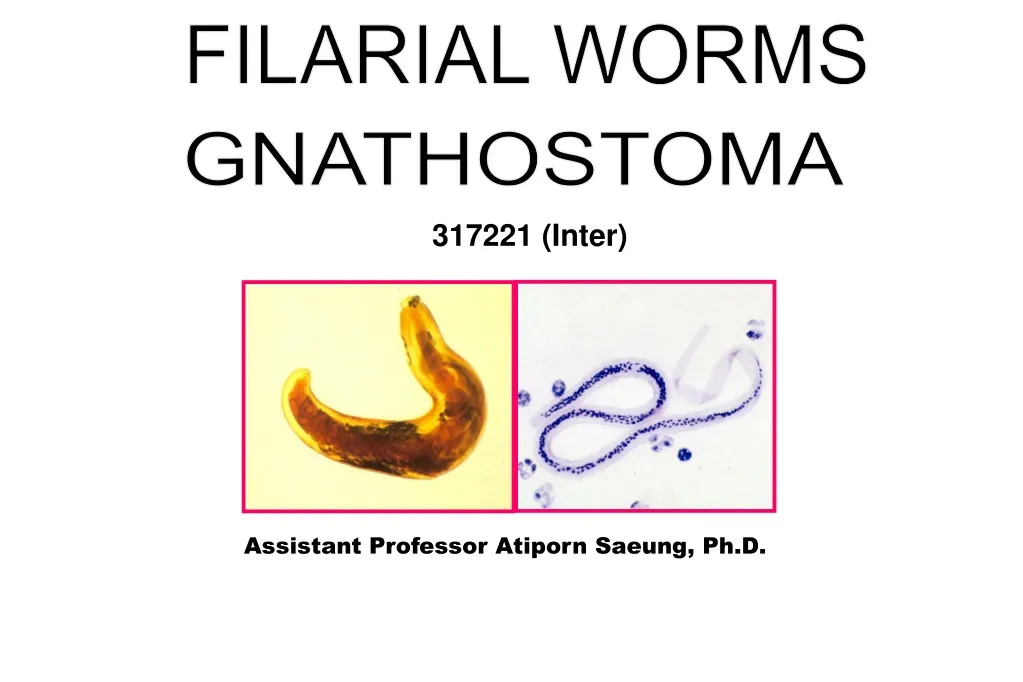
FILARIAL WORMS. GNATHOSTOMA. 317221 (Inter). Assistant Professor Atiporn Saeung , Ph.D. Objectives. After class, student are able to describe … Morphology of Wuchereria bancrofti , Brugia malayi and Gnathostoma spinigerum . Life cycle of these nematodes .

E N D
FILARIAL WORMS GNATHOSTOMA 317221 (Inter) Assistant Professor AtipornSaeung, Ph.D.
Objectives After class, student are able to describe… • Morphology of Wuchereriabancrofti, Brugiamalayi and Gnathostomaspinigerum. • Life cycle of these nematodes. • Mosquito vectors and endemic areas in Thailand for W. bancroftiand B. malayi. • Transmission, pathogenesis, clinical manifestation, diagnosis, treatment and prevention of these nematodes.
SPIRURIDA: SPIRURID NEMATODES • Gnathostomaspp. • Dracunculusmedinensis • Thelaziaspp. • Filarialworms
Filarialworms Superfamily: Filarioidea Habitat: Vertebrate host • lymphatic vessel • tissue • body cavity Disease: Filariasis
Lymphatic filariasis (elephantiasis) • Neglected tropical disease (NTD) • Filarial parasites are transmitted to humans through • mosquitoes • Threatened: 852 million people (52 countries) • Infection: 120 million people • Physical disability: 40 million people • About 60% of those infected live in the WHO South-East Asia Region & 40% in the African Region, and the remainder in other tropical areas • 90% of infections are caused by Wuchereriabancrofti and the remainder by Brugiaspp. http://www.filariasis.us/whoweare.html (WHO 2018)
Life cycle L3 Male Female Microfilaria
filariasis filariasis Disease • W. bancrofti Bancroftian • B. malayi Malayan
Endemicareas Wuchereriabancrofti Northern & Western parts Brugiamalayi Southern part
cephalicspace sheath columnnuclei tail Morphologyofmicrofilaria
Morphologyofmicrofilaria Brugiamalayi Wuchereriabancrofti Figure long& gracefulshape short& kinkingshape palesheath Giemsastain pinksheath
Morphologyofmicrofilaria sheath Cephalicspace long (L/W = 1/1) (L/W = 2/1) short Wuchereriabancrofti Brugiamalayi
Morphologyofmicrofilaria Wuchereriabancrofti Brugiamalayi Columnnuclei Disperse Overlap
Morphologyofmicrofilaria Wuchereriabancrofti Brugiamalayi Caudalnuclei Absent Present (2)
DIFFERENTIATION : MICROFILARIA Wuchereriabancrofti Brugiamalayi - Long& gracefulshape - Short & kinking shape - Giemsa: palesheath - Pinksheath - Disperse nuclei - Overlapping nuclei - Short cephalic space (1/1) - Long cephalic space (2/1) - No caudal nuclei - 2 caudalnuclei
Microfilarialperiodicity Periodic occurrence Continuous occurrence
MF(%) MF (%) 100 50 0 DAY DAY NIGHT DAY DAY DAY DAY NIGHT NIGHT DAY DAY Nocturnallyperiodic 06 12 18 24 06 12 18 Diurnallyperiodic Microfilarialperiodicity Periodicoccurrence (DP) (NP) 100 50 0 06 12 18 24 06 12 18
MF (%) 100 50 0 DAY DAY NIGHT NIGHT DAY DAY 06 12 18 24 06 12 18 Microfilarialperiodicity Continuous occurrence Nonperiodic
(DSP) MF(%) MF(%) 100 50 0 DAY DAY DAY DAY NIGHT NIGHT NIGHT NIGHT DAY DAY DAY DAY 06 12 18 24 06 12 18 Diurnallysubperiodic Microfilarialperiodicity Continuousoccurrence (NSP) 100 50 0 06 12 18 24 06 12 18 Nocturnallysubperiodic
Nocturnally periodic Nonperiodic Diurnally periodic Nocturnally subperiodic Diurnally subperiodic Microfilarialperiodicity 1. Periodic occurrence 2. Continuous occurrence
Mosquito vectors • W. bancrofti Nocturnallyperiodic (urban type): Culexquinquefasciatus Nocturnallysubperiodic (rural type): Anopheles spp. Aedes spp. Mansonia (Ma. dives) Manguin et al. (2010) • B. malayi Nocturnally subperiodic: Mansoniaspp. Diurnallysubperiodic: Coquillettidiacrassipes
Clinicalmanifestation I. Asymptomaticphase II. Inflammatory(acute) phase III. Obstructive phase
II. Inflammatory(acute) phase Lymphadenitis Lymphangitis Orchitis Epididymitis Elephantoidfever
III. Obstructive phase • Elephantiasis W. bancrofti groinregion arms& legs - hydrocele - chyluria legs B. malayi
Pathogenesis Adult worm (3-10 cm) & lymphatic vessel Lymphatic endothelium: hyperplasia Cell infiltration: lymphocyte, eosinophil, histocyte, epitheroid cell, giant cell & fibroblasts Fibrosis & lymph blockage Elephantiasis
Diagnosis • Physical findings, clinical history • Routine blood tests: microfilaria • - Fresh blood (Direct smear) • - Blood film: Thin/Thick (20–60 μl) of finger-prick blood • - Concentration techniques • : Knott’s techniques • : Membrane filtration technique • : Microhematocrittube technique • - Microfilarialprovocation method • PCR-microfilariae DNA can be detected in human blood and in mosquitoes
BrugiaRapidtest • Manufactured by ReszonDiagnostics • International (Selangor, Malaysia) • Detection of IgG4 antibody against • Brugia spp. in human blood samples • Detection of W. bancrofti antigen in human blood samples The AlereFilariasis Test Strip (FTS)
Treatment (mf rate > 0.6%) Mass treatment Once-yearly, single-dose, 2-drugtreatment albendazole(400 mg) + DEC (6 mg/kg) +ivermectin(150-200 µg/kg) albendazole (400 mg) Active community-level surveillance (mfrate < 0.6%) Selective treatment DEC (6 mg/kg): 2 consecutive days for 1 year
Treatment Supportivetreatment (disabilityperson) Rehabitation (soapwith2% glycerine) Surgery (Kondoleonoperation)
Prevention • Personalprotection • Controlofmosquitovectors • Masstreatment
Gnathostomaspinigerum • Common name: • พยาธิตัวจี๊ด (Thai) • Disease: Gnathostomiasis, neurognathostomiasis (NG) • Distribution: worldwide • Asia • In Thailand, >3,173 cases (Serodiagnosisrecords) during 1996-2005 • Australia • Latin America (Waikaguland Chamacho 2007; Wang et al. 2008; 2012; Eamsobhana 2014)
Gnathostoma species Asia • Gnathostomaspinigerum* (Thailand, Japan) • Gnathostomahispidum(China, Japan, Taiwan, the Philippines, Malaysia, Thailand and Vietnam) • Gnathostomadoloresi(Japan) • Gnathostomanipponicum(Japan) Latin America (Mexico, etc.) • Gnathostomabinucleatum (Jongthawin et al. 2015)
Morphology: adult worms • Roundworm morphology • Distinct: Head bulb equipped with hooks • 8 rows • Female size: 1-2 x 25-54 mm • Male size: 1-1.5 x 11-25 mm • With 2 spicules and curved tail
Male Female 2 spicules
Morphology: 3rd stage larva Small in size Early L3 (eL3) = 0.5 mm Advanced (aL3) = 3-4 mm Reproductive organ not developed Only 4 rows of cervical spines aL3
Morphology: Egg 38-40 x 65-70 µm Oval shape Colorless Apolar plug at one end eggshell One or two-cells stage
Life cycle Cats, Dogs, Tiger: Definitive hosts (harbor adult worm) Human: accidental host (aL3) Paratenic hosts (aL3) L1 in water Fish: 2nd intermediate hosts (eL3-> aL3) Cyclops: 1st intermediate hosts (L2-> eL3) Courtesy of link.sprinker.com
Transmission • Man acquires infection from: • Ingesting L2 or eL3-infected cyclops(unfiltered water from natural sources) • Consumption of raw meat (IH & PH) infected with aL3 • Penetration through the skin by aL3 • Transplacental migration
Pathogenesis • Migration of larvae in the body: • Tissue damage • Hemorrhage • Host response to worm secretion: • Protease (major): matrix metalloproteinases(invasion of host tissue) • Inflammatory responses to migrating larvae • Eosinophil, lymphocyte & plasma cell
Clinical manifestations Cutaneous gnathostomiasis • Intermittent migratory swelling hand, leg, face, eyelid, abdomen, etc. • Most common manifestation of gnathostomiasis • Incubation period: 3-4 weeks after infection • Itching • Creeping eruption (Cornaglia et al. 2016)
Visceral gnathostomiasis Ocular gnathostomiasis • Larval migration to eyelid/anterior chamber • Uveitis • Hemorrhage • Retinal scar & blindness
eye Visceral gnathostomiasis:
Clinical presentation of 248 patients with NG (Katchanovet al. 2011)
Visceral gnathostomiasis (cont.) • Pulmonary gnathostomiasis • cough, chest pain & hydropneumothorax • Gastrointestinal gnathostomiasis • Genitourinary gnathostomiasis
Diagnosis • Clinical diagnosis • History: consuming raw meat dishes made from fish, amphibians, reptiles, birds, or mammals • Signs and symptoms • CT/MRI in case of cerebral gnathostomiaisis • Laboratory examination: eosinophilia > 10% • CSF examination: xanthochromic/bloody • Pleocytosis with eosinophil > 30% • Normal glucose levels • Normal or elevated protein levels


