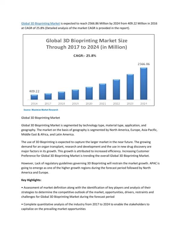
Download
1 / 11
130 likes | 731 Views
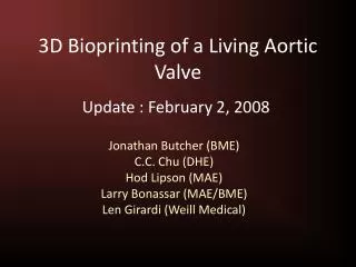
3D Bioprinting of a Living Aortic Valve. Update : February 2, 2008. Jonathan Butcher (BME) C.C. Chu (DHE) Hod Lipson (MAE) Larry Bonassar (MAE/BME) Len Girardi (Weill Medical). Clinical Need and State of the Art. Nearly 100,000 valve replacements annually in US

E N D
3D Bioprinting of a Living Aortic Valve Update : February 2, 2008 Jonathan Butcher (BME) C.C. Chu (DHE) Hod Lipson (MAE) Larry Bonassar (MAE/BME) Len Girardi (Weill Medical)
Clinical Need and State of the Art • Nearly 100,000 valve replacements annually in US • Prosthetic valves poor choice for young/active • Tissue engineering has potential but limited by inability to mimic 3D anatomy and heterogeneous material properties Native Aortic Valve Mechanical Properties TE Aortic Valve
Ideal Biomaterial Characteristics for Engineered Heart Valves • Enzymatically bioadsorbable • Cell mediated, non-toxic end products • Aqueous based hydrogel • Can fabricate with cells distributed within matrix • Non-thrombogenic/non-immunogenic • Tunable material properties: crosslinking • Bio-functionality • Charge, hydrophobicity, hydroxyl/amine groups
Arginine Based PEA Hydrogels (A-PEA) • Precursors are water soluble • Can be photo-crosslinked by UV light • Degraded by a variety of cellular enzymes • Numerous accessible functional groups WF68DA WF68DA/A2 WF68DA/A4 WF68DA/A3
Monocytes secreted over 5-fold less IL-6 on PEAs than on other polymers, 24 hrs A-PEA is Minimally Immunogenic/Thrombogenic IL-6 : proinflammatory cytokine, ↑ macrophage cytotoxic activity Monocytes on PEAs secreted less IL-1β, a potent pro-inflammatory cytokine, that can increase the surface thrombogenicity of the endothelium, 24 hrs MediVas TCT 04
3D Hydrogel Cytotoxicity Assay 96 well 3D gels with aortic valve interstitial cells
3D Cyotoxicity with Photo-Crosslinking 96 well 3D gels with aortic valve interstitial cells 90K cells/gel
Mechanical Testing of Hydrogels Grips Environmental Chamber Hydrogel Load Cell
High Throughput Measurement of Photo-Crosslinking Effects • Riboflavin induced crosslinking of collagen I • Central disk punched out via well guide • Dose dependent effects
Next Steps • Switch to A-PEA based hydrogels • Cytotoxicity of crosslinking dose • Mechanical testing of crosslinking effects • Incorporate a second syringe in the printer • Print a temporary “scaffold” to support structure • Print 3D anatomical models of heart valves • Axisymmetric aortic valve geometry • Anatomical models via MRI: Yi Wang, Weill Med • Incorporate a tuned UV laser to the print head • Spot specific engineered tissue material properties