Download
1 / 6
70 likes | 368 Views
Diestrous Phase. Stringy mucous, white blood cells (not many in this photo), nucleated epithelial cells. Proestrous. Mostly nucleated epithelial cells. Early Estrous. Relatively even distribution of nucleated epithelial cells and cornified epithelial cells. Estrous.

E N D
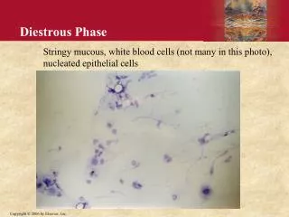
Diestrous Phase Stringy mucous, white blood cells (not many in this photo), nucleated epithelial cells
Proestrous Mostly nucleated epithelial cells
Early Estrous Relatively even distribution of nucleated epithelial cells and cornified epithelial cells
Estrous Mostly cornified epithelial cells
Metestrous Many WBC, some nucleated and cornified epithelial cells
Vaginal smear procedure: • Use clean pipet tip and inject about 25 microliters of 0.9% saline into the vagina of the rat. • With same tip, obtain a sample of the vaginal lavage and transfer to a clean slide (label with initials and the date). • Let dry, then stain for approximately 30-45 seconds in Wright stain (place a drop or two of stain on your slide). • Rinse slide briefly in water, then observe smear under microscope. Record description of smear and determine estrous phase. Retain slide for future reference if needed (store in labeled box in animal room).