acid base balance
Electrolyte and acid base balance


acid base balance
E N D
Presentation Transcript
Acid base balance Professor/mohammed Ahmed Bamashmos
Pathophysiology • Definition Of Acid-Base Balance: • This regulation of the extracellular fluid environment involves the ratio of acid to base, measured clinically as pH. • Physiologically all positively charged ions are called acids, and all negatively charged ions are bases. • Physiological changes in the concentration of H+ ions in the blood lead to acid-base balance. • A systemic increase in the H+ ions concentration is called acidosis. • A systemic decrease in the H+ ions is called alkalosis. • The acid-base must be regulated within a narrow range for the body to function normally. • A very slight change in the pH will affect the body.
H+ ions are needed for: • To maintain the integrity of the membrane. • Speed of the metabolic reactions. • Any change in the pH will lead to harmful effects than other diseases. • The symbol pH represents the power of H+. • When pH changes one unit like 7.0 to 6.0 = [H+] [H+] = H+ ions concentration changes 10 folds. • Body acids are formed from end products of: • Metabolism of proteins. • Metabolism of Carbohydrates. • Metabolism of fats. • This must be balanced by the number of basic substances in the body to maintain the normal pH. • Lungs, kidneys, and bones are the major organs involved in the regulation of acid-base balance
Body acids are formed from end products of: • Metabolism of proteins. • Metabolism of Carbohydrates. • Metabolism of fats. • This must be balanced by the number of basic substances in the body to maintain the normal pH. • Lungs, kidneys, and bones are the major organs involved in the regulation of acid-base balance. • Body acids are of two types: • Volatile acids: • Carbonic acid (H2CO3) is a week acid, and it does not easily release the H+ ions. • In the presence of carbonic anhydrase enzyme can eliminate CO 2 gas and water H2O. • CO 2 is eliminated through the lungs. • Nonvolatile acids: • These are sulfuric acid, phosphoric acid, and other organic acids that are eliminated through the kidneys. • These are the strong acids and readily give up their H+ ions. • Nonvolatile acids are secreted into the urine by the renal tubules. • These acids are about 150 meq/L of H+ ions per day or about 1 meq/kg body weight
Buffer Systems Of The Acid-Base Balance: • The buffer systems become active in response to change in the pH of the body as acid-base balance. • Functions ofthe buffer system: • Prevent the significant change in pH. • Buffer can absorb the excess of the H+ ions (acid). • Buffer system can absorb OH– ions, Hydroxyl (base). • The buffer system is present in the intracellular fluid (ICF) and extracellular fluid (ECF). • The most buffer system is: • Carbonic acid-bicarbonate system. • Hemoglobin system. • Phosphate and protein are the most important intracellular buffers (ICF
Renal buffering system:The distle tubule of the kidneys regulates acid-base balance by secreting the H+ ions in the urine and reabsorbs the HCO3–. • Dibasic phosphate (HPO4—) and ammonia (NH3) are two important renal buffer. • The renal buffering of H+ ions requires CO2 and water (H2O) to form the H2CO3. • The enzyme carbonic anhydrase catalyzes the reaction. • H+ ions are secreted from the tubular cells and buffer in the lumen by PO4— and NH3 = H2PO–3 + NH4+. • The rest of HCO3– is reabsorbed.
Carbonic acid-bicarbonate buffering system:This buffer system operates both in the lungs and kidneys. • This is the major extracellular buffer system. • Lungs can decrease the carbonic acid by blowing out the CO2 and leaving water behind. • Kidneys can reabsorb HCO3- or regenerate new HCO3- from CO 2 and water. • Normal bicarbonate (24 meq/L) and normal carbonic acid (1.2 meq/L), producing a 20:1 relation and maintain the pH of 7.4 • Both the systems are very efficient because: • HCO3– is easily reabsorbed or regenerated by the kidneys. • The lungs adjust acid concentration
Protein buffering system:Hemoglobin (Hb) is the best intracellular buffer system, and it combines with H+ and forming HHb and CO2, forming the HHbCO2 complex. • When Hb combines with H+ ions becomes weak acid. • Venous blood Hb is a better to buffer system than arterial blood Hb.
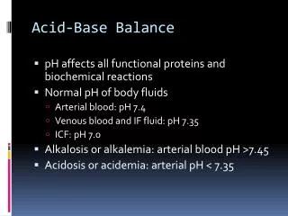
Acid-base balance: • H+ ions and electrolytes disturbances may be: • Acute. • Chronic. • Modest or severe. • Simple or mixed. • When there is an accumulation of H+ ions is called acidosis. • When blood pH is declining below 7.3, this process is called acidemia. • When there is a deficiency of H+ ions is called alkalosis. • Blood pH rises above 7.45 is called alkalemia. • There are conditions related to the respiratory system that leads to respiratory acidosis or alkalosis. • There are metabolic conditions related to kidneys, and abnormality of intake/output leads to metabolic acidosis/alkalosis.
The blood pH is normally maintained at 7.38 to 7.42. Any deviation from this range indicates a change in the H+ ions concentration. • Blood pH is a negative logarithm of [H+] as shown in the following equation: • pH = log10 [H+] • This equation shows that an increase in the H+ ions will lead to a fall in the blood pH is called acidemia. • So a decrease in the H+ ions will lead to an increase in the pH of the blood called alkalemia. • The conditions which cause the change in the pH are called acidosis and alkalosis. • The following diagram can explain how pH is maintained by the arterial carbon dioxide tension (pCO2) and plasma bicarbonate (HCO3–).
Plasma HCO3– decrease in the plasma caused by gastrointestinal or renal losses will increase H+ ions and lowers the pH.
Metabolic acidosis • Definition: • Metabolic acidosis occurs whenever there is a primary decrease in the HCO3¯ in the blood. • This may occur due to: • Exogenous acid administration. • Endogenous acid production. • Impaired renal H+ secretion. • HCO3– losses from the kidney or in the gastrointestinal secretions. • Anion gap: • Definition of the anion gap: • Anion gap referred to anions usually not measured in the laboratory like sulfate, phosphate, and lactate. The anions usually measured are Chloride (Cl-) and bicarbonate (HCO3-). The sum of the anions is subtracted from the sum of cations (Na+ and +); there is a gap around 10 to 12 meq/L, which is called an anion gap. An elevated anion gap gives clues for acidosis.
The importance of the anion gap is to identify the etiology of metabolic acidosis. • The anion gap is measured in meq/L. • Definition of anion gap: This is the difference between the plasma concentration of major cation sodium (Na+) and other anions are HCO3– and Cl–. • Anion gap = [Na+] – ([HCO3–] + [Cl–]) • The normal anion gap is 3 to 13 meq/L, and the mean is 10 meq/L. • This is dependant mainly on the plasma protein, primarily albumin. • 2.5 meq/L falls for every 1 gram/dl of albumin concentration in the blood. • H+ ions changes in the blood lead to acid-base imbalance. • A systemic increase in the H+ ions is called acidosis. • In the case of acidemia pH of the arterial blood is <7.4.
While in alkalemia, the pH of the arterial blood is >7.4.There is a systemic decrease in the H+ ions in the systemic blood is called alkalosis.
Causes Of Metabolic Acidosis: • In metabolic acidosis, noncarbonic acid increases, or HCO3¯ is lost from the extracellular space. • The buffering system becomes active and maintains the pH. • In case of the buffering system’s failure, the anion gap HCO3¯: H2CO3 = 20:1 changes. • Increased noncarbonic acid with an elevated anion gap and Increased H+ load: • Diabetes mellitus with ketoacidosis. There is a production of acetoacetic acid and β-hydroxybutyric acid in diabetic acidosis. • In the case of starvation. • Lactic acidosis in shock and hypoxemia. There is the production of lactic acid. • Ingestion of drugs like NH4CL, salicylates, methanol, ethylene glycol, and paraldehyde. • Decreased H+ ions excretion was seen in: • Uremia. • Distal renal tubular acidosis (decreased renal H+ secretion). • There is an accumulation of the acid that consumes the bicarbonate (HCO3¯).
Causes Of A High Anion Gap (>12 Meq/L): • Methanol toxicity. • Uremia due to renal failure. • Starvation. • Diabetes mellitus (ketoacidosis). • Lactic acidosis. • Salicylates toxicity. • Ethyl alcohol toxicity. • Isoniazid toxicity. • Iron toxicity. • Causes Of Decreased Anion Gap (<6 Meq/L): • Hypoalbuminemia. • Plasma cell disorders. • Bromide intoxication.
Causes Of A High Anion Gap (>12 Meq/L): • Methanol toxicity. • Uremia due to renal failure. • Starvation. • Diabetes mellitus (ketoacidosis). • Lactic acidosis. • Salicylates toxicity. • Ethyl alcohol toxicity. • Isoniazid toxicity. • Iron toxicity. • Causes Of Decreased Anion Gap (<6 Meq/L): • Hypoalbuminemia. • Plasma cell disorders. • Bromide intoxication.
Respiratory acidosis • Definition: • With respiratory failure, CO2 accumulates (hypercapnia). This state will raise the pCO2 and causes the pH to drop, and lead to acidosis. • This is a decrease in alveolar ventilation in relation to the metabolic production of the CO2 produces respiratory acidosis by the increase in the carbonic acid.
Pathophysiology:Alveolar ventilation provides the necessary oxygen for oxidative metabolism and eliminates the CO2 produced by these metabolic processes. • There is a depression in the ventilation, resulting in excess of CO2 (hypercapnia) in the blood circulation. • A decrease in alveolar ventilation in relation to the metabolic production of CO2 produces respiratory acidosis by an increase in H2CO3 acid. • The arterial CO2 tension (or pressure) PaCO2 is >45 mm Hg.
This is seen in respiratory failure, where CO2 accumulates, called hypercapnia. • This condition will raise the pCO2 and causes the pH to drop. • To compensate, the HCO3– will increase, but this is not sufficient to restore the pH to a normal level. • CO2 level rises, and this retained CO2 combines with water and form H2CO3. • H2CO3 dissociates to release H+ and HCO3– ions. • Increased paCO2 and free H+ ions stimulate the medulla to increase the respiratory rate and expel the CO2. • As the pH falls, 2.3, diphosphoglycerate accumulates in the RBCs, where it will alter the Hb (hemoglobin) to release the O2 (oxygen). • Hb picks up H+ ions and CO2 and removes both from the blood circulation. • If the respiratory mechanism fails, rising paCO2 stimulates the kidneys, retains HCO3– and Na+ (sodium) ions, and starts excreting H+ ions. • Total CO2 may rise to a very high level of chronic respiratory acidosis
Signs And Symptoms: • There is often breathlessness. • The patient is restless. • There is headache, dyspnoea, and tachypnea. • There is apprehension followed by lethargy. • The patient will have disorientation. • There are muscle twitching and tremors. • Skin will be warm and flushed due to raised CO2 causes vasodilatation. • There may be hypertension or hypotension. • There are atrial and ventricular arrhythmias. • The patient will have convulsions and ultimately goes into a coma.
Lab diagnosis: • pH = <7.35 to 7.45. • paCO2 = >45 mm Hg. • HCO3– = Normal (in the acute stage). • HCO3– = Increased (in the chronic stage
Treatment • Treatment of the pulmonary causes: • If there is obstruction by the foreign body, remove that immediately. • There may be a need for mechanical ventilators. • Give bronchodilators. • If there is pneumonia, then start antibiotics. • If there is pneumothorax, then put chest tube. • In the case of pulmonary embolism, start thrombolytic and anticoagulants. • Remove the secretions by bronchoscopy. • Treatment of chronic obstructive pulmonary disease (COPD): • Give O2 at a slow rate. • Start bronchodilators. • Start corticosteroids. • You can also give I/V sodium bicarbonate. • Other drugs are needed for the treatment of the cause.
cause. • Causes Of Respiratory Acidosis: • Acute respiratory acidosis: • This occurs with sudden obstruction to: • The airway. • Chest trauma that damages the respiratory muscles. • Acute paralysis or depression of CNS respiratory center. • HCO3– rises 1 meq/L for each 10 mmHg rise in pCO2. • Chronic respiratory acidosis: • This chronic respiratory acidosis is difficult to treat as compared to acute respiratory acidosis. • This will take place by: • Chronic obstructive pulmonary diseases like bronchitis, emphysema, pulmonary fibrosis, or scarring. • Accumulation of the CO2 lasting days, weeks, or months will provoke a sustained increase in HCO3– generation and leads to enhanced renal excretion of the H+ ions with chronic CO 2 retention. • HCO3– rises 3.5 meq/L for each 10 mm Hg rise in pCO 2. • The serum level of Na+ and K+ may be normal or mildly raised.
Suppression of the medullary respiratory center: • Sleep apnea. • Sedation medicines. • Cardiopulmonary arrest. • Upper respiratory obstruction: • Laryngospasm. • Aspiration of the foreign body or vomitus. • Obstruction in sleep apnea. • Defective respiratory muscle function: • Myasthenia gravis. • Guillain-barre syndrome. • Botulism. • Hypokalemia (severe). • Poliomyelitis. • Myxedema. • Amyotrophic lateral sclerosis. • Defect in the pulmonary gas exchange: • Acute respiratory distress syndrome. • Pneumothorax. • Hemothorax. • Severe asthma. • Severe pneumonia. • Chronic obstructive pulmonary disease
Respiratory Alkalosis • Definition: • This is due to over-breathing, causing excessive CO2 excretion, leading to a rise in blood pH. • Pathophysiology: • Overbreathing causes excessive CO2 exhaled out and causing the blood pH to rise.
Acute respiratory alkalosis interacts with intracellular and protein buffers before affecting the HCO3– system. • After the adjustment, blood HCO3– drops 5 meq/L for every 10 mmHg decline in pCO2. • Alkalosis causes plasma proteins to have a more negative charge that in turn binds more ionized Ca++. • This hypocalcemia increases neuromuscular excitability and leads to tetany. • Respiratory alkalosis occurs when there are alveolar hyperventilation and excessive reduction in plasma CO2 levels. This is called hypocapnia. • In the case of initial hypoxemia, there is increased ventilation mostly mediated by the chemoreceptors in the carotid body; these are located near the carotid artery’s bifurcation.
Kidneys compensate by decreasing H+ excretion and HCO3¯ reabsorption. • The PaCO2 is <35 mm Hg.
Causes Of Respiratory Alkalosis: • Pulmonary diseases due to hypoxemia: • Pneumonia. • Pulmonary embolism. • Pulmonary edema. • High-altitude syndrome. • Severe anemia. • Congestive heart failure. • Stimulation of the medullary (respiratory) center: • Hepatic encephalopathy. • Sepsis with fever. • Salicylates toxication. • Hyperventilation syndrome. • Pregnancy when there is increased progesterone. • Cerebrovascular accidents. • Pontine tumors.
Hypermetabolic conditions:Fever. • Anemia. • Thyrotoxicosis. • Hysteria. • Cirrhosis. • Gram-negative sepsis. • Pregnancy.
Signs And Symptoms: • The central and peripheral nervous system is stimulated, leading to: • There is light-headedness or Dizziness. • The patient may be agitated. • Confusion. • Tingling of the extremities appears first around the mouth and in the fingers and toes, called circumoral and peripheral paresthesia. • There is a carpopedal spasm, twitching, and muscle weakness. • Light-headedness and weakness may occur and progress to unconsciousness. • Convulsions. • Ultimately the patient goes into a coma. • Deep and rapid respirations are the primary symptoms that cause respiratory alkalosis. • Diagnosis: