Download

1 / 10
90 likes | 101 Views
quick view of Anatomy of respiratory system

E N D
Anatomy of respiratory system Prep. By : kyriolls Shaker Mahfouz
1 ILOS • INTRODUCTION • ANATOMY OF NOSE • ANATOMY OF PHARYNX • ANATOMY OF LARYNX • ANATOMY OF TRACHIA • ANATOMY OF LUNG
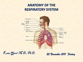
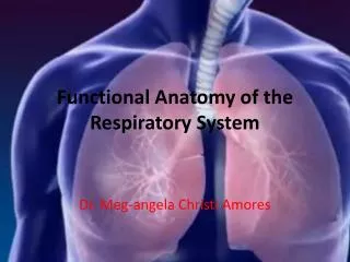
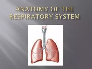
2 INTRODUCTION • The respiratory system is divided into two main portions : • Conducting portion : Nose, Pharynx, Larynx, Trachea and Bronchi • Respiratory portion Respiratory bronchioles, Alveolar ducts and Alveoli
3 Nose • Nasal cavity it’s a large, air-filled space above and behind the nose in the middle of the face. It’s divided into two cavities (fossae) by nasal septum. • Paranasal Sinuses air-filled space within the maxillary, frontal, ethmoid, and sphenoid bones of the skull
4 PHAYRNX • Thepharynxis the part of the throat behind the mouth and nasal cavity, and above the esophagus and trachea • It’s a common passageway for air and food • It’s composed of : • nasopharynx : uppermost portion • oropharynx : middle portion • laryngeopharynx: lowermost portion
5 Larynx • The larynx (voice box) is an organ located in the anterior neck. • The interior surface of it is lined bypseudostratified ciliated columnar epithelium(Respiratory epithelium) • the epiglottis which is An elastic cartilaginous flap, closing the laryngeal inlet prior to swallowing to prevent the food and liquid accessing the airways.
6 Trachea • The trachea is a cartilaginous tube that connects the larynx to the bronchi of the lungs, allowing the passage of air. • The interior surface of it is lined by pseudostratified ciliated columnar epithelium(Respiratory epithelium) • It is surrounded by 16 - 20 rings of hyaline cartilage incomplete and C-shaped, to keep it open and prevent collapsing
7 Bronchi • The bronchi are the airways that lead from the tracheainto the lungs and then branch off into smaller structures until they reach the alveoli. • They are made up of cartilage, smooth muscle, and mucous membranes. The trachea and the structures of the bronchi are known as the tracheobronchial tree, or the bronchial tree. • The right main bronchusis shorter and more vertical than the left, about 2.5 cm in length. • The left main bronchusis smaller and longer than the right main bronchus, about 5 cm. • They then divide into smaller branches to enter the lung ( 3 to the 3 lobes of the right lung, 2 to the 2 lobes of the left lung).
8 REFERENCES • https://en.m.wikipedia.org/wiki/Nasal_cavity#:~:text=The%20nasal%20cavity%20is%20a,one%20of%20the%20two%20nostrils.https://teachmeanatomy.info/neck/viscera/pharynx/ • https://teachmeanatomy.info/neck/viscera/larynx/organ/ • https://www.medicalnewstoday.com/articles/trachea. • https://www.verywellhealth.com/what-is-the-bronchus-structure-function-and-conditions-2249066#:~:text=The%20bronchi%20are%20the%20airways,carbon%20dioxide%20in%20the%20lungs.