Download
1 / 34
410 likes | 1.06k Views
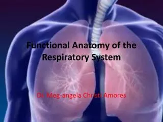
L-7 Respiratory System Functional Anatomy and Respiratory Volume. Dr Than Kyaw 26 March 2012. Topics (general outline). Introduction Functional anatomy Pulmonary ventilation Exchange of gases through pulmonary membrane V/Q ratio Regulation of respiration

E N D
L-7 Respiratory SystemFunctional Anatomy and Respiratory Volume Dr Than Kyaw 26 March 2012
Topics (general outline) • Introduction • Functional anatomy • Pulmonary ventilation • Exchange of gases through pulmonary membrane • V/Q ratio • Regulation of respiration • Factors affecting respiration rate • Hypoxia, dyspnea, and cynosis
Introduction • O2 - Vital requirement of animal • Animals may live - for days without water or - for weeks without food • Without O2 - live for a few minutes only! • This vital function -- done by respiratory system
Functions of respiratory system Primary functions - delivery of O2 on tissues - removal of CO2 (product of cellular respiration) - related function – ventilation Secondary functions (non-respiratory functions) - regulation of pH of body fluid (how) - thermoregulation (how) - phonation (making sounds) - defends against microbes (how) - removes some chemicals as well as producing others (how) - trap and dissolve blood clots (how) - smelling (olfactory epithelium – at the caudal portion of nasal cavities
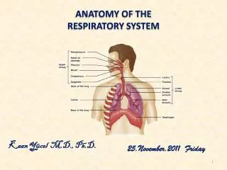
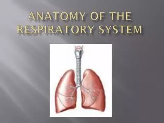
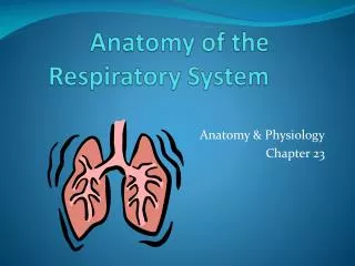
Respiratory apparatus • Air passages to lungs – nostrils -- nasal cavities, -- pharynx -- larynx -- trachea -- bronchi -- bronchioles -- alveoli • Lungs • Pleura
Air passages to lungs – nostrils (nares) -- external openings to the air passage -- Horse – most pliable(easily bends) and dilatable -- Pig – most rigid -- dilatability – advantageous when more air is required -- especially for horse (runner)
Air passages to lungs – nasal cavities -- paired and separated by nasal septum -- each consists of mucosa-covered turbinate bones (conchae); separating the nasal cavity into dorsal, middle and ventral meatus - cooling effect to blood supplying the brain (in the conchae - arteries supplying the brains divide many smaller artires and rejoin before entering the brain; as a result brain temperature: 2 – 3C lower than core body temperature.) -- mucosa - well vascularized - serve to warm and humidify inhaled air when more air is required -- especially for horse (runner)
Pharynx • Common passage way for air & food • Opennings to the pharynx -- 2 posterior nares -- 2 eustachian tubes -- oral cavity -- glottis -- esophagus • Larynx – organ of sound production in mammals Syrinx -- organ of sound in birds • Glottis -- slit-like opening ( site for endotracheal tubing) • Epiglottis – leaf-like extension from larynx, at the root of the tongue -- passively bend over larynx during the act of swallowing -- prevent bolus from entering the trachea
Cranial view of canineglottis, opening to the larynx between vocal cords and epiglottis.
Trachea • Continuation from the larynx • tracheal rings -- incomplete -- permits variation in diameter regulated by tracheal smooth m/s • diameter can increase during times of greater ventilatory requirements • Bronchi • Right and left • Bronchioles • Terminal bronchioles • Respiratory bronchioles • Alveolar duct • Alveolar sac • Alveoli
Pulmonary alveoli • Principle sites of gas exchange between the air and blood • Diffusion distance is minimal at alveolar level • Alveolar epithelium and capillary epithelium are intimately associated.
Alveolar cell types Alveolar type I cells. -- Squamous cells, as thin as 0.05 m; 95% of the alveolar epithelial surface. Alveolar type II cells. -- Irregular, cuboidal shaped; cytoplasm -- Cytosomes granules secrete pulmonary surfactant -- Surfactant -- protein-phospholipid mixture -- reduce the surface tension of the alveoli -- prevent collapse of alveoli during exhalation, and -- act as a bactericide
Features of Alveoli for efficient gas exchange • large surface area to absorb oxygen (about 70 Sq. meters in man). • moist surface to allow oxygen to dissolve. • thin lining to allow easy diffusion of gases ( >1 µ) • diameter - 7 • dense network of blood capillaries for easy gas exchange.
Features of capillaries for efficient gas exchange • dense network -- to carry CO2 and O2 • Large surface area to transport gases • Lining is one cell thick so gases can pass through quickly and easily. • Carbondioxide diffuses 20 times faster than oxygen • Change in thickness- Fibrosis • – affect gas exchange
Lungs and Pleura Lungs • pair; occupy all space in the thorax • Expansion of thorax provide air inflow into the lungs lungs expand • Air – radiolucent (penetrable by X-ray) -- air-filled lungs provide good contrast for thoracic structures that are radio-opaque • Blood – relatively radiopaque, can be seen in the X-ray
Lungs and Pleura Pleura • Serous membrane, friction-free movement of lungs • 2 layers – visceral (covering the lung) -- parietal (also k/s costal; attached to the thoracic wall - Intrapleural space – filled with fluid • Mediasternal space -- the junction of 2 pleural sacs near the midline of the thorax in which are found heart, vena cava, esophagus, thoracic lymph duct. • Pleuritis, pleurisy -- friction, difficult breathing, severe sharp pain
Respiration/Respiratory cycle 2 phases 1. Inspiration-- involves enlargement of thorax and lungs accompanied by air inflow Enlargement of thorax by contraction of diaphram and appropriate intercostal muscles. Inspiration need greater effort than expiration • Expiration – passive -- appropriate intercostal contraction -- abdominal m/s contraction -- force abdominal viscera forward to press on the diaphragm decrease thoracic vol.
2 types of breathing • Abdominal Breathing -- predominate in normal condition -- vissible abdominal contraction -- protrude during inspiration and recoil during expiration • Intercostal Breathing -- characterized by pronounced rib movements -- painful condition of abdominal (e.g peritonitis)
Muscles involved in Inhalation • Diaphragm: • -- contraction draws air into lungs • -- 75% of normal air movement • Externalintercostalmuscles: • -- assist inhalation by raising rib cage • -- 25% of normal air movement • Accessorymuscles assist in elevating ribs: • -- E.g. serratus , pectoralis, scalene muscles Lower Intrathoracic pressure
Muscles involved in exhalation • Internal intercostal and • transversusthoracis muscles: • -- depress the ribs • 2. Abdominal muscles: • -- compress the abdomen • -- force diaphragm upward increase Intrathoracic pressure
Terminology for States of breathing -- variations: -- frequency of respiratory cycle -- depth of inspiration -- both Eupnea-- normal quiet breathing Dyspnea -- difficulty breathing Hyperpnea -- depth & frequency – notable after physical exertion Polypnea -- rapid shallow breathing (panting) -- similar to hyperpnea in regard to frequency but not in depth Apnea -- transient cessation of breathing Tachypnea – excessive rapidity of breathing Bradypnea -- abnormal slowness of breathing
Pulmonary volumes and capacities Tidal volume -- amount of air breathed in or out during a respiratory cycle -- can increase or decrease from normal depending on ventilation requirement Inspiratory reserve vol. -- amount of air that can still be inspired after inhaling the tidal volume Expiratory reserve vol. -- amount of air that can still be expired after exhaling the tidal volume
Pulmonary volumes and capacities Residual vol -- the amount of air remaining in the lungs after the most forceful expiration Total lung capacity -- the sum of all volumes Vital capacity -- the difference between total volume and residual volume -- it is also the maximum amount of air that can be breathed in after the most forceful expiration
Pulmonary volumes and capacities Inspiratory capacity -- the sum of tidal and inspiratory reserve volume Functional Residual vol -- the sum of expiratory reserve volume and residual volume -- serve as reservoir for air and help to provide constancy to the blood concentration of the respired gas
Spirometer tracing showing the relationship between lung capacity and respiratory volume.
NOTE: Floating property of the lung Lungs of dead animals -- Because of remains of residual volume in the lung , excised lung sections of dead animal or slaughtered animals float in water Fetal lungs – consistency – like liver, no air, sink in water -- after birth and even one breath – residualair left -- the lung float in water due to residual air -- Determine whether newborn animal was born dead? Pneumonic lungs -- due to consolidation – lung tissue sinks in water