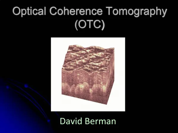
OPTICAL COHERENCE TOMOGRAPHY
Optical coherence tomography is all about modern analysis technique in pharmaceutical industry , optical as well as in coating technique

OPTICAL COHERENCE TOMOGRAPHY
E N D
Presentation Transcript
OPTICAL COHERENCE TOMOGRAPHY:PRINCIPLE AND APPLICATIONS RANJANA AHIRWAR M.Tech Pharma 2nd Semester DEPARTMENT OF PHARMACEUTICAL TECHNOLOGY (PROCESS CHEMISTRY) NIPER S.A.S NAGAR
FLOW OF PRESENTATION INTRODUCTION EVOLUTION PRINCIPLE TYPES OF OCT GENERATIONS OF OCT ADVANTAGES AND LIMITATIONS INSTRUMENTS APPLICATIONS CONCLUSION
OPTICAL COHERENCE TOMOGRAPHY • Optical : Relating or involving light and optics • Coherence: Two waves are coherent if they have a constant phase difference and same frequency • Tomography : Imaging by sectioning or slicing
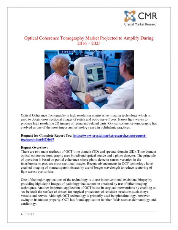
INTRODUCTION • OCT is noncontact, non invasive imaging technique used to obtain high resolution cross-sectional images of the retina and anterior segment • OCT allows the acquisition of cross-sections in a non-destructive and contactless manner • OCT is an imaging technique that uses coherent light to capture micrometer resolution, two- and three- dimensional images from within optical scattering media • It relies on exposing a sample to a burst of light and then measuring the reflective response from different depths and is therefore capable of scanning non-invasively beneath the surface of the sample • It is used for medical imaging and industrial non-destructive testing
OCT of the retina is like doing a vertical biopsy section of the retina. Instead of a knife, light is used. Instead of viewing a stained section under a microscope, we are presented with a “ false-colour” view with micron level resolution • There is no physical contact with the eye. OCT of the retina is the most important diagnostic tool for retinal pathology since the advent of fluorescein angiography
EVOLUTION • First, OCT techniques, like the reflectometry technique and the dual beam technique were based on time-domain low coherence interferometry • depth-scans • Later, Fourier-domain techniques have been developed and led to new imaging schemes • Recently developed parallel OCT schemes eliminate the need for lateral scanning and, therefore, dramatically increase the imaging rate. These schemes use CCD cameras and CMOS detector arrays as photodetectors • Modifying interference microscopy techniques has led to high-resolution optical coherence microscopy that achieved sub-micrometre resolution
QUALITATIVE AND QUANTITATIVE ANALYSIS • The OCT allows both qualitative and quantitative analysis of the retina • Qualitative analysis involves describing or identifying morphological changes and anomalous structures in the retina. Morphology is the study of forms and structures of organisms • Quantitative analysis is possible because the OCT software is able to identify and “trace” two key layers of the retina, the NFL and RPE. The software can then measure the distance between these two layers, which represents retinal thickness
RESOLUTION v/s PENETRATION Optical coherence tomography is an analogous to ultrasound, except that it uses light rather than sound. Unlike ultrasound, OCT does not require contact with the tissue examined
OCT v/s ULTRASOUND • OCT image has a resolving power of about 10µ vertically and 20µ horizontally • Compared that to the resolution of good ultrasound at 100µ • Ultrasound needs contact with tissue under study whereas OCT does not require any contact
PRINCIPLE • The principle of OCT is based on low coherence interferometry (Michelson interferometer) • Uses near infrared light (830-850nm) light coupled to a fibre optic system • In conventional interferometry with long coherence length (i.e,laser interferometry), interference of light occurs over a distance of meters • In OCT, this interference is shortened to a distance of micrometers, owing to the use of broad-bandwidth light sources (i.e, sources that emit light over a broad range of frequencies) • Light with broad bandwidths can be generated by using super luminescent diodes or lasers with extremely short pulses (femtosecond lasers).White light is an example of a broadband source with lower power
Contd… ►Near infrared light waves are partially reflected at different depths within the sample, and their arrival times at a detector are interferometrically compared with a reference wave ►The detected signal contains information on the position of the scatterers within the sample, based on their reflectivity , velocity and polarisation properties. Cross-sectional images can be reconstructed by collecting depth scans at different adjacent positions
Contd… • Interferometry is the technique of superimposing (interfering) two or more waves, to detect differences between them • Interferometry works because two waves with the same frequency that have the same phase will add each other while two waves that have opposite phase will subtract
Contd… • Light from a source is directed onto a partially reflecting mirror and is split into a reference and a measurement beam • The measurement beam reflected from the specimen with different time delays according to its internal microstructure
Contd… • The light in the reference beam is reflected from a reference mirror at a variable distance which produces a variable time delay • The light from the specimen, consisting of multipleechoes, and the light from the reference mirror, consisting of a single echo are travels back to the beam splitter and recombines to form interference pattern which is sensed and detected by photodetector
TYPES OF OCT 1. TIME DOMAIN OCT • In TD-OCT a mirror in the reference arm of the interferometer is moved to match the delay in various layers of the sample • The resulting interference is processed to produce the axial scan waveform • The reference mirror must move one cycle for each axial scan. The need for mechanical movement limits the speed of image acquisition • Further more, at each moment the detection system only collects signal from a narrow range of depth in the sample. This serial axial scanning is inefficient
FOURIER/SPECTRAL DOMAIN OCT • In FD-OCT the reference mirror is kept stationary. The spectral pattern of the interference between the sample and the reference reflections is measured • The spectral interferogram is fourier transformed to provide an axial scan. The absence of moving part allows the image to be acquired very rapidly • Furthermore, reflections from all layers in the sample are detected simultaneously. This parallel axial scan is much more efficient, resulting in both greater speed and higher signal-to-noise ratio
FD-OCT can be implemented in two ways: • In the swept-source implementation, a tunable laser is used to sequentially sweep through the spectrum, while the signal is collected by a single-element photodetector. This is called Swept-source OCT (SS-OCT) or optical Fourier domain imaging (OFDI) • In the spectrometer-based implementation, a broad-spectrum light source, such as a super luminescent diode (SLD), is used, and a spectrometer is utilized in the detector arm of the interferometer. The spectrometer uses a grating or a prism to spread the light into a spectrum. The spectrum is typically detected by a line camera. This technique has been called FD-OCT, spectral OCT spectral domain (SD) OCT, spectral RADAR, and frequency Domain (FD)
THE OCT MACHINE • The OCT system comprises: i) Fundus viewing unit ii) Interferometric unit iii) Computer display iv) Control panel v) Colour inkjet printer
GENERATIONS OF OCT • OCT 1, i.e, first generation of OCT machine has a transverse and axial resolution of 20 and 10 µ, respectively • OCT 2, i.e, second generation of OCT machine has a resolution similar to OCT 1 but with an improved user interface • Both OCT 1 and OCT 2 acquire 100 vertical scans in a standard OCT scan in an acquisition time of approximately 1.2 seconds • OCT 3, i.e, third-generation OCT unit has improved axial resolution of 7-8µ and acquires 512 vertical scans
Ophthalmology -diagnosing retinal diseases Dermatology -skin diseases Early detection of skin cancers Cardio-vascular diseases -vulnerable plaque detection Endoscopy (fibre-optic devices) -gastroenterology -gynecology Embryology/Developmental biology USE OF OCT IN MEDICAL SCIENCE Functional imaging -Doppler OCT (blood flow) -spectroscopic OCT (absorption,high speed) -optical properties - Polarization Sensitive-OCT(birefringence) • Guided surgery • Delicate procedures • Brain surgery • Knee surgery
ADVANTAGES OF OCT 1) Live sub-surface images at near-microscopic resolution 2) Instant, direct imaging of tissue morphology 3) No preparation of the sample or subject 4) No ionizing radiation LIMITATIONS The technique is limited to imaging 1 to 2 mm below the surface in biological tissue , because at greater depths the proportion of light that escapes without scattering is too small to be detected
INSTRUMENTS: 3D OCT-1 Maestro, Optical coherence tomography Simply touch the operation screen, and the maestro automatically scans both eyes and produces simultaneously an OCT scan and a true colour fundus image KEY FEATURES: 1) 50,000 A-scans per second 2) 12mm x 9mm wide field OCT scan for optic nerve and macula 3) True colour fundus image 4) Fully automated operation auto alignment, auto focus and auto shoot 5) Operation from any position 6) Widefield OCT
3D OCT-1 Maestro solo • Topcon 3D OCT-1 Maestro Solo incorporates an OCT for the posterior segment of the eye • KEY BENEFITS:- • Fully automated operation; auto alignment , auto focus & auto shoot • Operation from any position • Excellent interaction with patients • Space saving, due to small footprint • High definition B-scan composed of 50,000 A-scan per second • 12mm x 9mm wide field OCT scan for optic nerve and macula
CARL ZEISS Cirrus HD-OCT 5000 This OCT machine uses the Advanced RPE Analysis , providing highest resolution visualization capabilities and effectively tracking retinal pigment epithelial integrity The device features an active eye-trafficking mechanism which can trace and compensate eye motion in real time. Inbuilt FastTrac retinal tracking reduces eye motion artifacts without sacrificing the comfort of your patients
NIDEK OCT RS-3000 Advance Nidek OCT RS-3000 Advance is an economical OCT machine that is comprised of the main body which captures images and the computer which handles the processes and provides storage space The machine is perfect for evaluation of the choroid and the retina KEY FEATURES : 1) Retinal Camera and OCT combined 2) Anterior imaging 3) Superb macula image using SLO technology 4) Wide-field imaging from macula to disc 5) Tracing HD plus for accuracy 6) Selectable OCT sensitivity for enhanced visualization where required 7) Accurate image capture using the Torsion Eye Tracer (TET) technology 8) Multifunctional customized reports for easy follow-ups
OPTOVueRTVue Fourier Domain SD OCT System • Standing in the mid-price range, this OCT machine uses an innovative Fourier-Domain SD-OCT system • This innovative Fourier-Domain SD-OCT system enables the OptovueRTVue to perform high-speed, high-resolution tomography scanning that often used for retina imaging and analysis in clinical environments • OptovueRTVue works at extremely high clarity, which means only a fraction of a second is required to visualize the retinal tissue and evaluate post-refractive patients accurately, providing a realistic prognosis
Contd… • KEY FEATURES:- • Ultra-high speed and high-density mapping • High resolution • Two scan depth for imaging a tilted disk • Two image modes • High transverse scan resolution • Video fundus image for high-quality retina image • Non-mydriatic imaging for fundus imaging
CONCLUSION Optical coherence tomography is a valuable clinical tool for the diagnosis and management of retinal diseases and glaucoma. Its use has enable better understanding, diagnosis, decision making and management in a wide variety of conditions. Major technological advances in OCT are certain to impact research and clinical practice in ophthalmology in the future.